

BAX Primary Antibody
Item Information
Catalog #
Size
Price
Description
The protein encoded by this gene belongs to the BCL2 protein family. BCL2 family members form hetero- or homodimers and act as anti- or pro-apoptotic regulators that are involved in a wide variety of cellular activities. This protein forms a heterodimer with BCL2, and functions as an apoptotic activator. This protein is reported to interact with, and increase the opening of, the mitochondrial voltage-dependent anion channel (VDAC), which leads to the loss in membrane potential and the release of cytochrome c. The expression of this gene is regulated by the tumor suppressor P53 and has been shown to be involved in P53-mediated apoptosis. Multiple alternatively spliced transcript variants, which encode different isoforms, have been reported for this gene.
Product Overview
Entrez GenelD
581
Aliases
BCL2L4
Clone#
4H9B11
Host / Isotype
Mouse / IgG2a
Species Reactivity
Human
Immunogen
Purified recombinant fragment of human BAX (AA: 13-160) expressed in E. Coli.
Formulation
Purified antibody in PBS with 0.05% sodium azide
Storage
Store at 4°C short term. Aliquot and store at -20°C long term. Avoid freeze/thaw cycles.
Product Applications
WB (Western Blot)
1/500 - 1/2000
IHC_P(Immunohistochemistry)
1/200 - 1/1000
ELISA
1/10000
References
1.Rev Esp Enferm Dig. 2015 Jul;107(8):520-1.
2.Cell Death Dis. 2015 Jul 9;6:e1809.
2.Cell Death Dis. 2015 Jul 9;6:e1809.
Product Image
Elisa
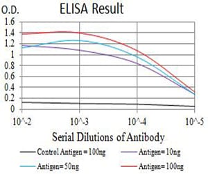
Figure 1: Black line: Control Antigen (100 ng);Purple line: Antigen (10ng); Blue line: Antigen (50 ng); Red line:Antigen (100 ng)
Western Blot
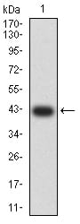
Figure 2:Western blot analysis using BAX mAb against human BAX (AA: 13-160) recombinant protein. (Expected MW is 42.5 kDa)
Western Blot
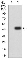
Figure 3:Western blot analysis using BAX mAb against HEK293 (1) and BAX (AA: 13-160)-hIgGFc transfected HEK293 (2) cell lysate.
Immunohistochemical analysis
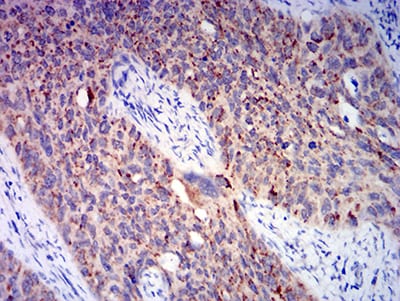
Figure 4:Immunohistochemical analysis of paraffin-embedded cervical cancer tissues using BAX mouse mAb with DAB staining.
Immunohistochemical analysis
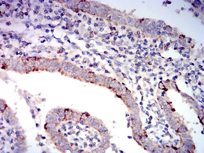
Figure 5:Immunohistochemical analysis of paraffin-embedded endometrial cancer tissues using BAX mouse mAb with DAB staining.
For Research Use Only. Not for use in diagnostic procedures.