

BAD Primary Antibody
Item Information
Catalog #
Size
Price
Description
The protein encoded by this gene is a member of the BCL-2 family. BCL-2 family members are known to be regulators of programmed cell death. This protein positively regulates cell apoptosis by forming heterodimers with BCL-xL and BCL-2, and reversing their death repressor activity. Proapoptotic activity of this protein is regulated through its phosphorylation. Protein kinases AKT and MAP kinase, as well as protein phosphatase calcineurin were found to be involved in the regulation of this protein. Alternative splicing of this gene results in two transcript variants which encode the same isoform.
Product Overview
Entrez GenelD
572
Aliases
BBC2; BCL2L8
Clone#
1G5B3
Host / Isotype
Mouse / IgG1
Species Reactivity
Human
Immunogen
Purified recombinant fragment of human BAD (AA: FULL(1-168)) expressed in E. Coli.
Formulation
Purified antibody in PBS with 0.05% sodium azide.
Storage
Store at 4°C short term. Aliquot and store at -20°C long term. Avoid freeze/thaw cycles.
Product Applications
WB (Western Blot)
1/500 - 1/2000
FCM (Flow Cytometry)
1/200 - 1/400
ELISA
1/10000
References
1. Clin Cancer Res. 2011 Oct 1;17(19):6356-66.
2. Mol Cancer Res. 2010 Aug;8(8):1116-25.
2. Mol Cancer Res. 2010 Aug;8(8):1116-25.
Product Image
Western Blot
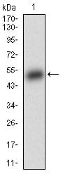
Figure 1: Western blot analysis using BAD mAb against human BAD (AA: FULL(1-168)) recombinant protein. (Expected MW is 44.3 kDa)
Western Blot
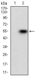
Figure 2: Western blot analysis using BAD mAb against HEK293 (1) and BAD (AA: FULL(1-168))-hIgGFc transfected HEK293 (2) cell lysate.
Western Blot

Figure 3: Western blot analysis using BAD mouse mAb against MCF-7 (1), HEK293 (2) cell lysate.
Flow cytometric

Figure 4: Flow cytometric analysis of MCF-7 cells using BAD mouse mAb (green) and negative control (red).
Elisa
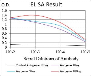
Black line: Control Antigen (100 ng); Purple line: Antigen(10ng); Blue line: Antigen (50 ng); Red line: Antigen (100 ng);
For Research Use Only. Not for use in diagnostic procedures.