

Mouse Monoclonal Antibody to B4GALNT1
Item Information
Catalog #
Size
Price
Description
GM2 and GD2 gangliosides are sialic acid-containing glycosphingolipids. GalNAc-T is the enzyme involved in the biosynthesis of G(M2) and G(D2) glycosphingolipids. GalNAc-T catalyzes the transfer of GalNAc into G(M3) and G(D3) by a beta-1,4 linkage, resulting in the synthesis of G(M2) and G(D2), respectively. Three transcript variants encoding different isoforms have been found for this gene.
Product Overview
Entrez GenelD
2583
Aliases
GALGT; SPG26; GALNACT; GalNAc-T
Clone#
4G11B1
Host / Isotype
Mouse / IgG1
Immunogen
Purified recombinant fragment of human B4GALNT1 (AA: 26-225) expressed in E. Coli.
Formulation
Purified antibody in PBS with 0.05% sodium azide
Storage
Store at 4°C short term. Aliquot and store at -20°C long term. Avoid freeze/thaw cycles.
Product Applications
WB (Western Blot)
1/500 - 1/2000
IHC_P(Immunohistochemistry)
1/200 - 1/1000
FCM (Flow Cytometry)
1/200 - 1/400
ELISA
1/10000
References
1,Exp Mol Pathol. 2019 Aug;109:25-35. 2,Sci Rep. 2020 Jan 27;10(1):1199.
Product Image
Elisa

Figure 1:Black line: Control Antigen (100 ng);Purple line: Antigen (10ng); Blue line: Antigen (50 ng); Red line:Antigen (100 ng)
Western Blot
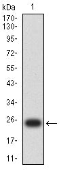
Figure 2:Western blot analysis using B4GALNT1 mAb against human B4GALNT1 (AA: 26-225) recombinant protein. (Expected MW is 24.4 kDa)
Western Blot

Figure 3:Western blot analysis using B4GALNT1 mAb against HEK293-6e (1) and human B4GALNT1 (AA: 26-225)-hIgGFc transfected HEK293-6e (2) cell lysate.
Western Blot

Figure 4:Western blot analysis using B4GALNT1 mouse mAb against Hela (1), Jurkat (2), HepG2 (3), k562 (4), COS-7 (5), and NIH/3T3 (6) cell lysate.
Flow cytometric analysis

Figure 5:Flow cytometric analysis of Hela cells using B4GALNT1 mouse mAb (green) and negative control (red).
Immunohistochemical analysis

Figure 6:Immunohistochemical analysis of paraffin-embedded brain tissues using B4GALNT1 mouse mAb with DAB staining.
Immunohistochemical analysis

Figure 7:Immunohistochemical analysis of paraffin-embedded cervical cancer tissues using B4GALNT1 mouse mAb with DAB staining.
For Research Use Only. Not for use in diagnostic procedures.