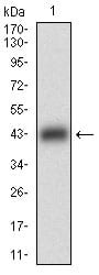
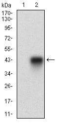
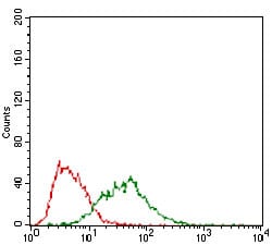
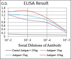
B3GALT4 Primary Antibody

Item Information
Catalog #
Size
Price
Description

This gene is a member of the beta-1,3-galactosyltransferase (beta3GalT) gene family. This family encodes type II membrane-bound glycoproteins with diverse enzymatic functions using different donor substrates (UDP-galactose and UDP-N-acetylglucosamine) and different acceptor sugars (N-acetylglucosamine, galactose, N-acetylgalactosamine). The beta3GalT genes are distantly related to the Drosophila Brainiac gene and have the protein coding sequence contained in a single exon. The beta3GalT proteins also contain conserved sequences not found in the beta4GalT or alpha3GalT proteins. The carbohydrate chains synthesized by these enzymes are designated as type 1, whereas beta4GalT enzymes synthesize type 2 carbohydrate chains. The ratio of type 1:type 2 chains changes during embryogenesis. By sequence similarity, the beta3GalT genes fall into at least two groups: beta3GalT4 and 4 other beta3GalT genes (beta3GalT1-3, beta3GalT5). This gene is oriented telomere to centromere in close proximity to the ribosomal protein S18 gene. The functionality of the encoded protein is limited to ganglioseries glycolipid biosynthesis.

Product Overview
Entrez GenelD
8705
Aliases
GALT2; GALT4; BETA3GALT4
Clone#
5F12B8
Host / Isotype
Mouse / IgG2a
Species Reactivity
Human
Immunogen
Purified recombinant fragment of human B3GALT4 (AA: 191-359) expressed in E. Coli.
Formulation
Purified antibody in PBS with 0.05% sodium azide.
Storage
4°C; -20°C for long term storage
Product Applications
WB (Western Blot)
1/500 - 1/2000
FCM (Flow Cytometry)
1/200 - 1/400
ELISA
1/10000
References
1. Immunogenetics. 2000 Jan;51(1):75-8.
2. Tumour Biol. 2009;30(1):43-50.
Product Image
Western Blot
Figure 1: Western blot analysis using B3GALT4 mAb against human B3GALT4 (AA: 191-359) recombinant protein. (Expected MW is 44.3 kDa)
Western Blot
Figure 2: Western blot analysis using B3GALT4 mAb against HEK293 (1) and B3GALT4 (AA: 191-359)-hIgGFc transfected HEK293 (2) cell lysate.
Western Blot
Figure 3: Western blot analysis using B3GALT4 mouse mAb against PANC-1 (1), PC-3 (2) cell lysate.
Flow cytometric
Figure 4: Flow cytometric analysis of PANC-1 cells using B3GALT4 mouse mAb (green) and negative control (red).
Elisa
Black line: Control Antigen (100 ng); Purple line: Antigen(10ng); Blue line: Antigen (50 ng); Red line: Antigen (100 ng);
For Research Use Only. Not for use in diagnostic procedures.