

B2M Primary Antibody
Item Information
Catalog #
Size
Price
Description
This gene encodes a serum protein found in association with the major histocompatibility complex (MHC) class I heavy chain on the surface of nearly all nucleated cells. The protein has a predominantly beta-pleated sheet structure that can form amyloid fibrils in some pathological conditions. A mutation in this gene has been shown to result in hypercatabolic hypoproteinemia.
Product Overview
Entrez GenelD
567
Clone#
3G5H8
Host / Isotype
Mouse / IgG2a
Species Reactivity
Human
Immunogen
Purified recombinant fragment of human B2M (AA: 21-100) expressed in E. Coli.
Formulation
Purified antibody in PBS with 0.05% sodium azide.
Storage
Store at 4°C short term. Aliquot and store at -20°C long term. Avoid freeze/thaw cycles.
Product Applications
WB (Western Blot)
1/500 - 1/2000
IHC_P(Immunohistochemistry)
1/200 - 1/1000
FCM (Flow Cytometry)
1/200 - 1/400
ELISA
1/10000
References
1. Cancer Immunol Immunother. 2012 Sep;61(9):1359-71.
2. Lupus. 2012 Sep;21(10):1098-104.
2. Lupus. 2012 Sep;21(10):1098-104.
Product Image
Western Blot

Figure 1: Western blot analysis using B2M mAb against human B2M (AA: 21-100) recombinant protein. (Expected MW is 35.4 kDa)
Western Blot

Figure 2: Western blot analysis using B2M mAb against HEK293 (1) and B2M (AA: 21-100)-hIgGFc transfected HEK293 (2) cell lysate.
Western Blot
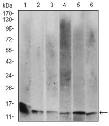
Figure 3: Western blot analysis using B2M mouse mAb against Hela (1), HEK293 (2), HepG2 (3),RAJI (4), A431 (5) and Jurkat (6) cell lysate.
Flow cytometric

Figure 4: Flow cytometric analysis of A431 cells using B2M mouse mAb (green) and negative control (red).
Immunohistochemical analysis

Figure 5: Immunohistochemical analysis of paraffin-embedded ovarian cancer tissues using B2M mouse mAb with DAB staining.
Immunohistochemical analysis

Figure 6: Immunohistochemical analysis of paraffin-embedded esophageal cancer tissues using B2M mouse mAb with DAB staining.
Elisa

Black line: Control Antigen (100 ng); Purple line: Antigen(10ng); Blue line: Antigen (50 ng); Red line: Antigen (100 ng);
For Research Use Only. Not for use in diagnostic procedures.