

AXIN1 Primary Antibody
Item Information
Catalog #
Size
Price
Description
This gene encodes a cytoplasmic protein which contains a regulation of G-protein signaling (RGS) domain and a dishevelled and axin (DIX) domain. The encoded protein interacts with adenomatosis polyposis coli, catenin beta-1, glycogen synthase kinase 3 beta, protein phosphate 2, and itself. This protein functions as a negative regulator of the wingless-type MMTV integration site family, member 1 (WNT) signaling pathway and can induce apoptosis. The crystal structure of a portion of this protein, alone and in a complex with other proteins, has been resolved. Mutations in this gene have been associated with hepatocellular carcinoma, hepatoblastomas, ovarian endometriod adenocarcinomas, and medullablastomas. Alternative splicing results in multiple transcript variants.
Product Overview
Entrez GenelD
8312
Aliases
AXIN; PPP1R49
Clone#
4E9F1
Host / Isotype
Mouse / IgG2b
Species Reactivity
Human
Immunogen
Purified recombinant fragment of human AXIN1 (AA: 546-752) expressed in E. Coli.
Formulation
Purified antibody in PBS with 0.05% sodium azide
Storage
Store at 4°C short term. Aliquot and store at -20°C long term. Avoid freeze/thaw cycles.
Product Applications
WB (Western Blot)
1/500 - 1/2000
ICC (Immunocytochemistry)
1/50 - 1/250
FCM (Flow Cytometry)
1/200 - 1/400
ELISA
1/10000
References
1.Cancer Lett. 2014 Dec 1;355(1):1-8.
2.BMC Cancer. 2013 Aug 2;13:368.
2.BMC Cancer. 2013 Aug 2;13:368.
Product Image
Elisa
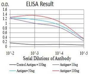
Figure 1: Black line: Control Antigen (100 ng);Purple line: Antigen (10ng); Blue line: Antigen (50 ng); Red line:Antigen (100 ng)
Western Blot

Figure 2:Western blot analysis using AXIN1 mAb against human AXIN1 (AA: 546-752) recombinant protein. (Expected MW is 48.7 kDa)
Western Blot

Figure 3:Western blot analysis using AXIN1 mAb against HEK293 (1) and AXIN1 (AA: 546-752)-hIgGFc transfected HEK293 (2) cell lysate.
Immunofluorescence analysis

Figure 4:Immunofluorescence analysis of Hela cells using AXIN1 mouse mAb (green). Blue: DRAQ5 fluorescent DNA dye. Red: Actin filaments have been labeled with Alexa Fluor- 555 phalloidin. Secondary antibody from Fisher (Cat#: 35503)
Flow cytometric

Figure 5:Flow cytometric analysis of Hela cells using AXIN1 mouse mAb (green) and negative control (red).
For Research Use Only. Not for use in diagnostic procedures.