

AURKA Primary Antibody
Item Information
Catalog #
Size
Price
Description
The protein encoded by this gene is a cell cycle-regulated kinase that appears to be involved in microtubule formation and/or stabilization at the spindle pole during chromosome segregation. The encoded protein is found at the centrosome in interphase cells and at the spindle poles in mitosis. This gene may play a role in tumor development and progression. A processed pseudogene of this gene has been found on chromosome 1, and an unprocessed pseudogene has been found on chromosome 10. Multiple transcript variants encoding the same protein have been found for this gene.
Product Overview
Entrez GenelD
6790
Aliases
AIK; ARK1; AURA; BTAK; STK6; STK7; STK15; PPP1R47
Clone#
2D8A4
Host / Isotype
Mouse / IgG2b
Species Reactivity
Human
Immunogen
Purified recombinant fragment of human AURKA (AA: 268-404) expressed in E. Coli.
Formulation
Purified antibody in PBS with 0.05% sodium azide
Storage
Store at 4°C short term. Aliquot and store at -20°C long term. Avoid freeze/thaw cycles.
Product Applications
WB (Western Blot)
1/500 - 1/2000
IHC_P(Immunohistochemistry)
1/200 - 1/1000
ICC (Immunocytochemistry)
1/200 - 1/1000
FCM (Flow Cytometry)
1/200 - 1/400
ELISA
1/10000
References
1.Mol Cancer Ther. 2015 Dec;14(12):2753-61.
2.Oncol Res Treat. 2015;38(9):442-7.
2.Oncol Res Treat. 2015;38(9):442-7.
Product Image
Elisa

Figure 1: Black line: Control Antigen (100 ng);Purple line: Antigen (10ng); Blue line: Antigen (50 ng); Red line:Antigen (100 ng)
Western Blot
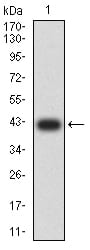
Figure 2:Western blot analysis using AURKA mAb against human AURKA (AA: 268-404) recombinant protein. (Expected MW is 41.5 kDa)
Western Blot

Figure 3:Western blot analysis using AURKA mAb against HEK293 (1) and AURKA (AA: 268-404)-hIgGFc transfected HEK293 (2) cell lysate.
Western Blot

Figure 4:Western blot analysis using AURKA mouse mAb against HEK293 (1), MCF-7 (2), and Hela (3) cell lysate.
Immunofluorescence analysis

Figure 5:Immunofluorescence analysis of Hela cells using AURKA mouse mAb (green). Blue: DRAQ5 fluorescent DNA dye. Red: Actin filaments have been labeled with Alexa Fluor- 555 phalloidin. Secondary antibody from Fisher (Cat#: 35503)
Immunofluorescence analysis

Figure 6:Immunofluorescence analysis of SMMC-7721 cells using AURKA mouse mAb (green). Blue: DRAQ5 fluorescent DNA dye. Red: Actin filaments have been labeled with Alexa Fluor- 555 phalloidin. Secondary antibody from Fisher (Cat#: 35503)
Flow cytometric

Figure 7:Flow cytometric analysis of HeLa cells using AURKA mouse mAb (green) and negative control (red).
Immunohistochemical analysis

Figure 8:Immunohistochemical analysis of paraffin-embedded cervical cancer tissues using AURKA mouse mAb with DAB staining.
Immunohistochemical analysis

Figure 9:Immunohistochemical analysis of paraffin-embedded rectum cancer tissues using AURKA mouse mAb with DAB staining.
For Research Use Only. Not for use in diagnostic procedures.