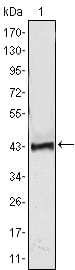
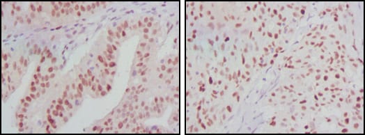
ATP2C1 Primary Antibody

Item Information
Catalog #
Size
Price
Description

ATP2C1, also known as PMR1, it belongs to the family of P-type cation transport ATPases. This magnesium-dependent enzyme catalyzes the hydrolysis of ATP coupled with the transport of the calcium. The human homologue, ATP2C1 (also designated SPLA in rat), also regulates the transport of calcium in the Golgi complex and is related to other P-type ATPases family members, such as the sarco(endo)plasmic calcium ATPase (SERCA) and the plasma membrane calcium ATPase (PCMA). ATP2C1 is a transmembrane protein that exists as two splice variants, which vary by 20 amino acids. Defects in ATP2C1 cause Hailey-Hailey disease, which is an autosomal dominant disorder that is characterized by blisters and erosions of the skin. These findings provide further evidence that PMR1 plays a key role in maintaining the integrity of the epidermis by controlling intracellular calcium signaling.

Product Overview
Entrez GenelD
27032
Aliases
HHD; BCPM; PMR1; SPCA1
Clone#
4G12
Host / Isotype
Mouse / IgG1
Species Reactivity
Human, Monkey
Immunogen
Purified recombinant fragment of ATP2C1 expressed in E. Coli.
Formulation
Ascitic fluid containing 0.03% sodium azide.
Storage
4°C; -20°C for long term storage
Product Applications
WB (Western Blot)
1/500 - 1/2000
IHC_P(Immunohistochemistry)
1/200 - 1/1000
ELISA
1/10000
References
1. J Invest Dermatol. 2005 Nov;125(5):933-5.
2. J Dermatol Sci. 2006 Aug;43(2):150-1.
3. Dermatology. 2007;215(4):277-83.
Product Image
Western Blot
Figure 1: Western blot analysis using ATP2C1 mAb against human ATP2C1 (AA: 119-269) recombinant protein. (Expected MW is 41.7 kDa)
Western Blot
Figure 2: Western blot analysis using ATP2C1 mouse mAb against A431 (1), Hela (2) and HEK293 (3) cell lysate.
Immunohistochemical analysis
Figure 3: Immunohistochemical analysis of paraffin-embedded human ovarian cancer (left) and breast cancer (right) tissues using ATP2C1 mouse mAb with DAB staining.
For Research Use Only. Not for use in diagnostic procedures.