
ATP2A1 Primary Antibody
Item Information
Catalog #
Size
Price
Description
This gene encodes one of the SERCA Ca(2+)-ATPases, which are intracellular pumps located in the sarcoplasmic or endoplasmic reticula of muscle cells. This enzyme catalyzes the hydrolysis of ATP coupled with the translocation of calcium from the cytosol to the sarcoplasmic reticulum lumen, and is involved in muscular excitation and contraction. Mutations in this gene cause some autosomal recessive forms of Brody disease, characterized by increasing impairment of muscular relaxation during exercise. Alternative splicing results in three transcript variants encoding different isoforms.
Product Overview
Entrez GenelD
487
Aliases
ATP2A; SERCA1
Clone#
3D1H6
Host / Isotype
Mouse / IgG1
Species Reactivity
Human, Mouse, Monkey
Immunogen
Purified recombinant fragment of human ATP2A1 (AA: 487-631) expressed in E. Coli.
Formulation
Purified antibody in PBS with 0.05% sodium azide
Storage
Store at 4°C short term. Aliquot and store at -20°C long term. Avoid freeze/thaw cycles.
Product Applications
WB (Western Blot)
1/500 - 1/2000
FCM (Flow Cytometry)
1/200 - 1/400
ELISA
1/10000
References
1.Biochem Biophys Res Commun. 2012 Jun 29;423(2):212-7.
2.Mol Genet Metab. 2013 Sep-Oct;110(1-2):162-9.
2.Mol Genet Metab. 2013 Sep-Oct;110(1-2):162-9.
Product Image
Elisa
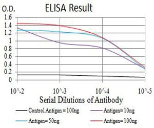
Figure 1: Black line: Control Antigen (100 ng);Purple line: Antigen (10ng); Blue line: Antigen (50 ng); Red line:Antigen (100 ng)
Western Blot
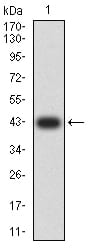
Figure 2:Western blot analysis using ATP2A1 mAb against human ATP2A1 (AA: 487-631) recombinant protein. (Expected MW is 42 kDa)
Western Blot
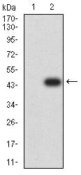
Figure 3:Western blot analysis using ATP2A1 mAb against HEK293 (1) and ATP2A1 (AA: 487-631)-hIgGFc transfected HEK293 (2) cell lysate.
Western Blot

Figure 4:Western blot analysis using ATP2A1 mouse mAb against C2C12 (1), COS7 (2), Hela (3), K562 (4), and Jurkat (5) cell lysate.
Flow cytometric
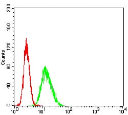
Figure 5:Flow cytometric analysis of HeLa cells using ATP2A1 mouse mAb (green) and negative control (red).
For Research Use Only. Not for use in diagnostic procedures.