

ATG5 Primary Antibody
Item Information
Catalog #
Size
Price
Description
The protein encoded by this gene, in combination with autophagy protein 12, functions as an E1-like activating enzyme in a ubiquitin-like conjugating system. The encoded protein is involved in several cellular processes, including autophagic vesicle formation, mitochondrial quality control after oxidative damage, negative regulation of the innate antiviral immune response, lymphocyte development and proliferation, MHC II antigen presentation, adipocyte differentiation, and apoptosis. Several transcript variants encoding different protein isoforms have been found for this gene.
Product Overview
Entrez GenelD
9474
Aliases
ASP; APG5; APG5L; hAPG5; APG5-LIKE
Clone#
3C10B6
Host / Isotype
Mouse / IgG2b
Species Reactivity
Human
Immunogen
Purified recombinant fragment of human ATG5 (AA: 144-275) expressed in E. Coli.
Formulation
Purified antibody in PBS with 0.05% sodium azide
Storage
Store at 4°C short term. Aliquot and store at -20°C long term. Avoid freeze/thaw cycles.
Product Applications
WB (Western Blot)
1/500 - 1/2000
FCM (Flow Cytometry)
1/200 - 1/400
ELISA
1/10000
References
1.PLoS One. 2014 Oct 17;9(10):e110293.
2.Sci Transl Med. 2013 Sep 11;5(202):202ra123.
2.Sci Transl Med. 2013 Sep 11;5(202):202ra123.
Product Image
Elisa

Figure 1: Black line: Control Antigen (100 ng);Purple line: Antigen (10ng); Blue line: Antigen (50 ng); Red line:Antigen (100 ng)
Western Blot

Figure 2:Western blot analysis using ATG5 mAb against human ATG5 (AA: 144-275) recombinant protein. (Expected MW is 41.5 kDa)
Western Blot
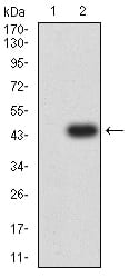
Figure 3:Western blot analysis using ATG5 mAb against HEK293 (1) and ATG5 (AA: 144-275)-hIgGFc transfected HEK293 (2) cell lysate.
Flow cytometric
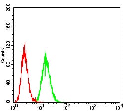
Figure 4:Flow cytometric analysis of Hela cells using ATG5 mouse mAb (green) and negative control (red).
For Research Use Only. Not for use in diagnostic procedures.