

ATG5 Primary Antibody
Item Information
Catalog #
Size
Price
Description
ATG5 involved in autophagic vesicle formation. Conjugation with ATG12, through a ubiquitin-like conjugating
system involving ATG7 as an E1-like activating enzyme and ATG10 as an E2-like conjugating enzyme, is essential
for its function. The ATG12-ATG5 conjugate acts as an E3-like enzyme which is required for lipidation of ATG8
family proteins and their association to the vesicle membranes. Involved in mitochondrial quality control after
oxidative damage, and in subsequent cellular longevity. The ATG12-ATG5 conjugate also negatively regulates the
innate antiviral immune response by blocking the type I IFN production pathway through direct association with
RARRES3 and MAVS. Also plays a role in translation or delivery of incoming viral RNA to the translation
apparatus. Plays a critical role in multiple aspects of lymphocyte development and is essential for both B and T
lymphocyte survival and proliferation. Required for optimal processing and presentation of antigens for MHC II.
Involved in the maintenance of axon morphology and membrane structures, as well as in normal adipocyte
differentiation. Promotes primary ciliogenesis through removal of OFD1 from centriolar satellites and degradation
of IFT20 via the autophagic pathway
Product Overview
Entrez GenelD
9474
Aliases
ASP; APG5; APG5L; hAPG5; APG5-LIKE
Clone#
8E8G6
Host / Isotype
Mouse / IgG2a
Species Reactivity
Human
Immunogen
Synthesized peptide of human ATG5 (AA: MTDDKDVLRDVWFGRIc).
Formulation
Purified antibody in PBS with 0.05% sodium azide
Storage
Store at 4°C short term. Aliquot and store at -20°C long term. Avoid freeze/thaw cycles.
Product Applications
WB (Western Blot)
1/500 - 1/2000
ICC (Immunocytochemistry)
1/200 - 1/1000
FCM (Flow Cytometry)
1/200 - 1/400
ELISA
1/10000
References
Autophagy. 2013 Jan;9(1):20-32.
Anticancer Res. 2012 Sep;32(9):4091-6.
Anticancer Res. 2012 Sep;32(9):4091-6.
Product Image
Elisa

Figure 1: Black line: Control Antigen (100 ng); Purple line: Antigen(10ng); Blue line: Antigen (50 ng); Red line: Antigen (100 ng);
Western Blot

Figure 2:Western blot analysis using ATG5 mouse mAb against Hela (1) and K562 (2) cell lysate.
Flow cytometric

Figure 3:Flow cytometric analysis of Hela cells using ATG5 mouse mAb (green) and negative control (red).
Immunofluorescence analysis
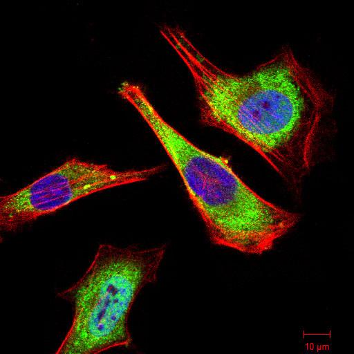
Figure 3:Immunofluorescence analysis of Hela cells using ATG5 mouse mAb (green). Blue: DRAQ5 fluorescent DNA dye. Red: Actin filaments have been labeled with Alexa Fluor- 555 phalloidin. Secondary antibody from Fisher (Cat#: 35503)
For Research Use Only. Not for use in diagnostic procedures.