

ATG4A Primary Antibody
Item Information
Catalog #
Size
Price
Description
Autophagy is the process by which endogenous proteins and damaged organelles are destroyed intracellularly. Autophagy is postulated to be essential for cell homeostasis and cell remodeling during differentiation, metamorphosis, non-apoptotic cell death, and aging. Reduced levels of autophagy have been described in some malignant tumors, and a role for autophagy in controlling the unregulated cell growth linked to cancer has been proposed. This gene encodes a member of the autophagin protein family. The encoded protein is also designated as a member of the C-54 family of cysteine proteases.
Product Overview
Entrez GenelD
115201
Aliases
APG4A; AUTL2
Clone#
8E8G2
Host / Isotype
Mouse / IgG1
Species Reactivity
Human
Immunogen
Purified recombinant fragment of human ATG4A (AA: 258-398) expressed in E. Coli.
Formulation
Purified antibody in PBS with 0.05% sodium azide
Storage
Store at 4°C short term. Aliquot and store at -20°C long term. Avoid freeze/thaw cycles.
Product Applications
WB (Western Blot)
1/500 - 1/2000
FCM (Flow Cytometry)
1/200 - 1/400
ELISA
1/10000
References
1.Breast Cancer Res. 2013 Nov 14;15(6):R109.
2.Autophagy. 2013 Jun 1;9(6):881-93.
2.Autophagy. 2013 Jun 1;9(6):881-93.
Product Image
Elisa
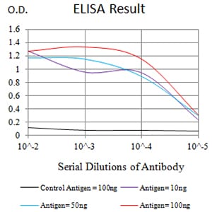
Figure 1:Black line: Control Antigen (100 ng);Purple line: Antigen (10ng); Blue line: Antigen (50 ng); Red line:Antigen (100 ng)
Western Blot

Figure 2:Western blot analysis using ATG4A mAb against human ATG4A (AA: 258-398) recombinant protein. (Expected MW is 42.2 kDa)
Western Blot

Figure 3:Western blot analysis using ATG4A mAb against HEK293 (1) and ATG4A (AA: 258-398)-hIgGFc transfected HEK293 (2) cell lysate.
Flow cytometric

Figure 4:Flow cytometric analysis of K562 cells using ATG4A mouse mAb (green) and negative control (red).
For Research Use Only. Not for use in diagnostic procedures.