
ATF4 Primary Antibody
Item Information
Catalog #
Size
Price
Description
This gene encodes a transcription factor that was originally identified as a widely expressed mammalian DNA binding protein that could bind a tax-responsive enhancer element in the LTR of HTLV-1. The encoded protein was also isolated and characterized as the cAMP-response element binding protein 2 (CREB-2). The protein encoded by this gene belongs to a family of DNA-binding proteins that includes the AP-1 family of transcription factors, cAMP-response element binding proteins (CREBs) and CREB-like proteins. These transcription factors share a leucine zipper region that is involved in protein-protein interactions, located C-terminal to a stretch of basic amino acids that functions as a DNA binding domain. Two alternative transcripts encoding the same protein have been described. Two pseudogenes are located on the X chromosome at q28 in a region containing a large inverted duplication.
Product Overview
Entrez GenelD
468
Aliases
CREB2; TXREB; CREB-2; TAXREB67
Clone#
2A6F12
Host / Isotype
Mouse / IgG1
Species Reactivity
Human
Immunogen
Purified recombinant fragment of human ATF4 (AA: 212-351) expressed in E. Coli.
Formulation
Purified antibody in PBS with 0.05% sodium azide
Storage
4°C; -20°C for long term storage
Product Applications
WB (Western Blot)
1/500 - 1/2000
IHC_P(Immunohistochemistry)
1/200 - 1/1000
ELISA
1/10000
References
1.Cell. 2014 Aug 28;158(5):1159-72.
2.Tumour Biol. 2014 Jan;35(1):765-71.
2.Tumour Biol. 2014 Jan;35(1):765-71.
Product Image
Elisa
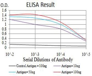
Figure 1: Black line: Control Antigen (100 ng);Purple line: Antigen (10ng); Blue line: Antigen (50 ng); Red line:Antigen (100 ng)
Western Blot
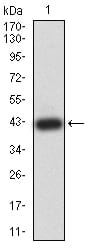
Figure 2:Western blot analysis using ATF4 mAb against human ATF4 (AA: 212-351) recombinant protein. (Expected MW is 41.5 kDa)
Western Blot
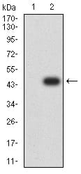
Figure 3:Western blot analysis using ATF4 mAb against HEK293 (1) and ATF4 (AA: 212-351)-hIgGFc transfected HEK293 (2) cell lysate.
Western Blot
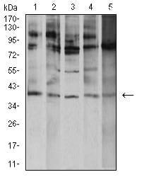
Figure 4:Western blot analysis using ATF4 mouse mAb against K562 (1), A431 (2), Hela (3), HEK293 (4), and Ramos (5) cell lysate.
Immunohistochemical analysis
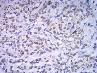
Figure 5:Immunohistochemical analysis of paraffin-embedded breast cancer tissues using ATF4 mouse mAb with DAB staining.
Immunohistochemical analysis
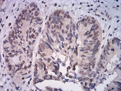
Figure 6:Immunohistochemical analysis of paraffin-embedded rectum cancer tissues using ATF4 mouse mAb with DAB staining.
For Research Use Only. Not for use in diagnostic procedures.