

ASS1 Primary Antibody
Item Information
Catalog #
Size
Price
Description
The protein encoded by this gene catalyzes the penultimate step of the arginine biosynthetic pathway. There are approximately 10 to 14 copies of this gene including the pseudogenes scattered across the human genome, among which the one located on chromosome 9 appears to be the only functional gene for argininosuccinate synthetase. Mutations in the chromosome 9 copy of ASS cause citrullinemia. Two transcript variants encoding the same protein have been found for this gene.
Product Overview
Entrez GenelD
445
Aliases
ASS; CTLN1
Clone#
2C10
Host / Isotype
Mouse / IgG1
Species Reactivity
Human, Mouse, Monkey
Immunogen
Purified recombinant fragment of human ASS1 expressed in E. Coli.
Formulation
Purified antibody in PBS with 0.05% sodium azide
Storage
Store at 4°C short term. Aliquot and store at -20°C long term. Avoid freeze/thaw cycles.
Product Applications
WB (Western Blot)
1/500 - 1/2000
IHC_P(Immunohistochemistry)
1/200 - 1/1000
ELISA
1/10000
References
1. Int J Cancer. 2009 Sep 15;125(6):1454-63.
2. Clin Biochem. 2009 Jul;42(10-11):1166-8.
2. Clin Biochem. 2009 Jul;42(10-11):1166-8.
Product Image
Western Blot

Figure 1: Western blot analysis using ASS1 mAb against human ASS1 (AA: 40-236) recombinant protein. (Expected MW is 47 kDa)
Western Blot
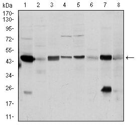
Figure 2: Western blot analysis using ASS1 mouse mAb against A431 (1), RAJI (2), MOLT4 (3), Jurkat (4), A549 (5), NIH/3T3 (6), PC-12 (7) and Cos7 (8) cell lysate.
Immunohistochemical analysis

Figure 3: Immunohistochemical analysis of paraffin-embedded cervical cancer tissues using ASS1 mouse mAb with DAB staining.
Immunohistochemical analysis

Figure 4: Immunohistochemical analysis of paraffin-embedded colon cancer tissues using ASS1 mouse mAb with DAB staining.
Elisa

Black line: Control Antigen (100 ng); Purple line: Antigen(10ng); Blue line: Antigen (50 ng); Red line: Antigen (100 ng);
For Research Use Only. Not for use in diagnostic procedures.