
ASH2L Primary Antibody
Item Information
Catalog #
Size
Price
Description
ASH2L (ASH2 Like Histone Lysine Methyltransferase Complex Subunit) is a Protein Coding gene. Diseases associated with ASH2L include Kabuki Syndrome 1. Among its related pathways are Chromatin organization and Signaling by GPCR. GO annotations related to this gene include transcription regulatory region DNA binding and histone methyltransferase activity (H3-K4 specific).
Product Overview
Entrez GenelD
9070
Aliases
ASH2; Bre2; ASH2L1; ASH2L2
Clone#
6B10H10
Host / Isotype
Mouse / IgG1
Species Reactivity
Human
Immunogen
Purified recombinant fragment of human ASH2L (AA: 493-628) expressed in E. Coli.
Formulation
Purified antibody in PBS with 0.05% sodium azide
Storage
Store at 4°C short term. Aliquot and store at -20°C long term. Avoid freeze/thaw cycles.
Product Applications
WB (Western Blot)
1/500 - 1/2000
IHC_P(Immunohistochemistry)
1/200 - 1/1000
ELISA
1/10000
References
1.Mol Cell. 2013 Mar 28;49(6):1108-20.
2.Nat Struct Mol Biol. 2011 Jun 5;18(7):857-9.
2.Nat Struct Mol Biol. 2011 Jun 5;18(7):857-9.
Product Image
Elisa
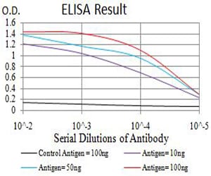
Figure 1: Black line: Control Antigen (100 ng);Purple line: Antigen (10ng); Blue line: Antigen (50 ng); Red line:Antigen (100 ng)
Western Blot
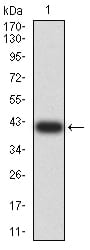
Figure 2:Western blot analysis using ASH2L mAb against human ASH2L (AA: 493-628) recombinant protein. (Expected MW is 41.6 kDa)
Western Blot
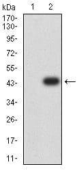
Figure 3:Western blot analysis using ASH2L mAb against HEK293 (1) and ASH2L (AA: 493-628)-hIgGFc transfected HEK293 (2) cell lysate.
Immunohistochemical analysis
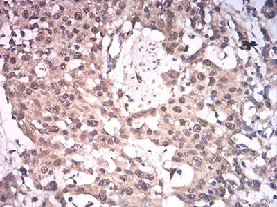
Figure 4:Immunohistochemical analysis of paraffin-embedded esophageal cancer tissues using ASH2L mouse mAb with DAB staining.
Immunohistochemical analysis
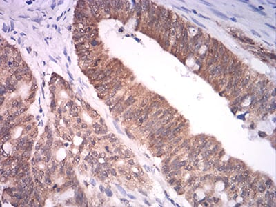
Figure 5:Immunohistochemical analysis of paraffin-embedded rectum cancer tissues using ASH2L mouse mAb with DAB staining.
For Research Use Only. Not for use in diagnostic procedures.