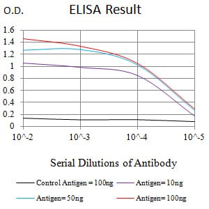
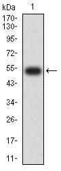
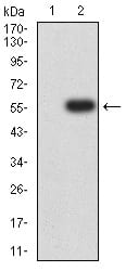
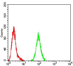
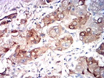
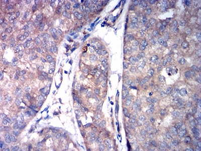
ASGR2 Primary Antibody

Item Information
Catalog #
Size
Price
Description

This gene encodes a subunit of the asialoglycoprotein receptor. This receptor is a transmembrane protein that plays a critical role in serum glycoprotein homeostasis by mediating the endocytosis and lysosomal degradation of glycoproteins with exposed terminal galactose or N-acetylgalactosamine residues. The asialoglycoprotein receptor may facilitate hepatic infection by multiple viruses including hepatitis B, and is also a target for liver-specific drug delivery. The asialoglycoprotein receptor is a hetero-oligomeric protein composed of major and minor subunits, which are encoded by different genes. The protein encoded by this gene is the less abundant minor subunit. Alternatively spliced transcript variants encoding multiple isoforms have been observed for this gene.

Product Overview
Entrez GenelD
433
Aliases
HL-2; HBXBP; ASGPR2; ASGP-R2; CLEC4H2
Clone#
2C6D11
Host / Isotype
Mouse / Mouse IgG1
Immunogen
Purified recombinant fragment of human ASGR2 (AA: 80-311) expressed in E. Coli.
Formulation
Purified antibody in PBS with 0.05% sodium azide
Storage
4?; -20? for long term storage
Product Applications
WB (Western Blot)
1/500 - 1/2000
IHC_P(Immunohistochemistry)
1/200 - 1/1000
FCM (Flow Cytometry)
1/200 - 1/400
ELISA
1/10000
References
1.Mol Cancer Res. 2018 Sep;16(9):1420-1429. 2.PLoS One. 2011;6(11):e27210.
Product Image
Elisa
Figure 1:Black line: Control Antigen (100 ng);Purple line: Antigen (10ng); Blue line: Antigen (50 ng); Red line:Antigen (100 ng)
Western Blot
Figure 2:Western blot analysis using ASGR2 mAb against human ASGR2 (AA: 80-311) recombinant protein. (Expected MW is 52.5 kDa)
Western Blot
Figure 3:Western blot analysis using ASGR2 mAb against HEK293 (1) and ASGR2 (AA: 80-311)-hIgGFc transfected HEK293 (2) cell lysate.
Flow cytometric
Figure 4:Flow cytometric analysis of Hela cells using ASGR2 mouse mAb (green) and negative control (red).
Immunohistochemical analysis
Figure 5:Immunohistochemical analysis of paraffin-embedded liver cancer tissues using ASGR2 mouse mAb with DAB staining.
Immunohistochemical analysis
Figure 6:Immunohistochemical analysis of paraffin-embedded bladder cancer tissues using ASGR2 mouse mAb with DAB staining.
For Research Use Only. Not for use in diagnostic procedures.