
ASF1B Primary Antibody
Item Information
Catalog #
Size
Price
Description
This gene encodes a member of the H3/H4 family of histone chaperone proteins and is similar to the anti-silencing function-1 gene in yeast. The encoded protein is the substrate of the tousled-like kinase family of cell cycle-regulated kinases, and may play a key role in modulating the nucleosome structure of chromatin by ensuring a constant supply of histones at sites of nucleosome assembly.
Product Overview
Entrez GenelD
55723
Aliases
CIA-II
Clone#
6G7G4
Host / Isotype
Mouse / IgG1
Species Reactivity
Human, Mouse, Monkey
Immunogen
Purified recombinant fragment of human ASF1B (AA: 1-202) expressed in E. Coli.
Formulation
Purified antibody in PBS with 0.05% sodium azide
Storage
Store at 4°C short term. Aliquot and store at -20°C long term. Avoid freeze/thaw cycles.
Product Applications
WB (Western Blot)
1/500 - 1/2000
FCM (Flow Cytometry)
1/200 - 1/400
ELISA
1/10000
References
1.EMBO J. 2011 Feb 2;30(3):480-93.
2.Mol Cell Biol. 2008 Jun;28(11):3672-85.
2.Mol Cell Biol. 2008 Jun;28(11):3672-85.
Product Image
Elisa
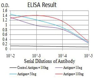
Figure 1: Black line: Control Antigen (100 ng);Purple line: Antigen (10ng); Blue line: Antigen (50 ng); Red line:Antigen (100 ng)
Western Blot
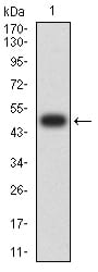
Figure 2:Western blot analysis using ASF1B mAb against human ASF1B (AA: 1-202) recombinant protein. (Expected MW is 48.4 kDa)
Western Blot
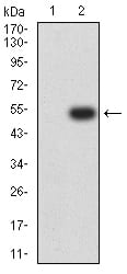
Figure 3:Western blot analysis using ASF1B mAb against HEK293 (1) and ASF1B (AA: 1-202)-hIgGFc transfected HEK293 (2) cell lysate.
Western Blot
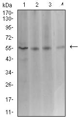
Figure 4:Western blot analysis using ASF1B mouse mAb against Hela (1), COS7 (2), HCT116 (3), and CHO3D10 (4) cell lysate.
Flow cytometric
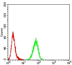
Figure 5:Flow cytometric analysis of K562 cells using ASF1B mouse mAb (green) and negative control (red).
For Research Use Only. Not for use in diagnostic procedures.