

ARG1 Primary Antibody
Item Information
Catalog #
Size
Price
Description
Arginase catalyzes the hydrolysis of arginine to ornithine and urea. At least two isoforms of mammalian arginase exist (types I and II) which differ in their tissue distribution, subcellular localization, immunologic crossreactivity and physiologic function. The type I isoform encoded by this gene, is a cytosolic enzyme and expressed predominantly in the liver as a component of the urea cycle. Inherited deficiency of this enzyme results in argininemia, an autosomal recessive disorder characterized by hyperammonemia. Two transcript variants encoding different isoforms have been found for this gene. [provided by RefSeq, Sep 2011]
Product Overview
Entrez GenelD
383
Aliases
ARG1
Clone#
6B4B11
Host / Isotype
Mouse / Mouse IgG1
Immunogen
Purified recombinant fragment of human ARG1 (AA: (1-322)) expressed in E. Coli.
Formulation
Purified antibody in PBS with 0.05% sodium azide
Storage
Store at 4°C short term. Aliquot and store at -20°C long term. Avoid freeze/thaw cycles.
Product Applications
WB (Western Blot)
1/500 - 1/2000
IHC_P(Immunohistochemistry)
1/200-1/1000
ELISA
1/10000
References
1,Medicine (Baltimore). 2020 Aug 7;99(32):e21634.2,Medicine (Baltimore). 2019 Nov;98(47):e17694.
Product Image
ELISA
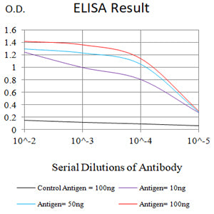
Figure 1: Black line: Control Antigen (100 ng);Purple line: Antigen (10ng); Blue line: Antigen (50 ng); Red line: Antigen (100 ng)
WESTERN BLOT
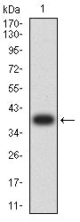
Figure 2: Western blot analysis using ARG1 mAb against human ARG1 (AA: (1-322)) recombinant protein. (Expected MW is 38.6 kDa)
WESTERN BLOT
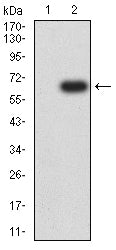
Figure 3: Western blot analysis using ARG1 mAb against HEK293-6e (1) and ARG1 (AA: (1-322))-hIgGFc transfected HEK293-6e (2) cell lysate.
IMMUNOHISTOCHEMISTRY
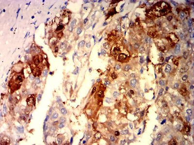
Figure 4: Immunohistochemical analysis of paraffin-embedded liver cancer tissues using ARG1 mouse mAb with DAB staining.
IMMUNOHISTOCHEMISTRY
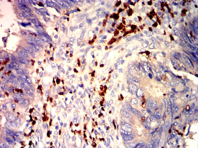
Figure 5: Immunohistochemical analysis of paraffin-embedded rectal cancer tissues using ARG1 mouse mAb with DAB staining.
For Research Use Only. Not for use in diagnostic procedures.