
ARF1 Primary Antibody
Item Information
Catalog #
Size
Price
Description
ADP-ribosylation factor 1 (ARF1) is a member of the human ARF gene family. The family members encode small guanine nucleotide-binding proteins that stimulate the ADP-ribosyltransferase activity of cholera toxin and play a role in vesicular trafficking as activators of phospholipase D. The gene products, including 6 ARF proteins and 11 ARF-like proteins, constitute a family of the RAS superfamily. The ARF proteins are categorized as class I (ARF1, ARF2 and ARF3), class II (ARF4 and ARF5) and class III (ARF6), and members of each class share a common gene organization. The ARF1 protein is localized to the Golgi apparatus and has a central role in intra-Golgi transport. Multiple alternatively spliced transcript variants encoding the same protein have been found for this gene.
Product Overview
Entrez GenelD
375
Aliases
N
Clone#
8D8E9
Host / Isotype
Mouse / IgG2a
Species Reactivity
Human
Immunogen
Purified recombinant fragment of human ARF1 (AA: 76-182) expressed in E. Coli.
Formulation
Purified antibody in PBS with 0.05% sodium azide
Storage
Store at 4°C short term. Aliquot and store at -20°C long term. Avoid freeze/thaw cycles.
Product Applications
WB (Western Blot)
1/500 - 1/2000
IHC_P(Immunohistochemistry)
1/200 - 1/1000
ICC (Immunocytochemistry)
1/200 - 1/1000
FCM (Flow Cytometry)
1/200 - 1/400
ELISA
1/10000
References
1.Mol Biol Cell. 2014 Jan;25(1):17-29.
2.Cancer Sci. 2012 Jun;103(6):1136-44.
2.Cancer Sci. 2012 Jun;103(6):1136-44.
Product Image
Elisa
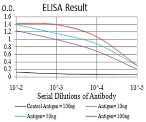
Figure 1: Black line: Control Antigen (100 ng);Purple line: Antigen (10ng); Blue line: Antigen (50 ng); Red line:Antigen (100 ng)
Western Blot
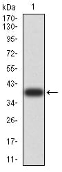
Figure 2:Western blot analysis using ARF1 mAb against human ARF1 (AA: 76-182) recombinant protein. (Expected MW is 39.3 kDa)
Western Blot
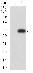
Figure 3:Western blot analysis using ARF1 mAb against HEK293 (1) and ARF1 (AA: 1-182)-hIgGFc transfected HEK293 (2) cell lysate.
Immunofluorescence analysis
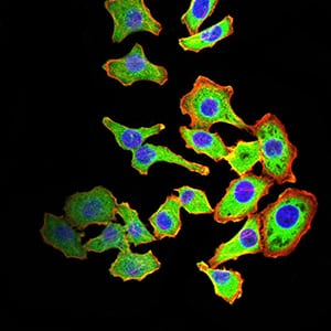
Figure 4:Immunofluorescence analysis of HL-7702 cells using ARF1 mouse mAb (green). Blue: DRAQ5 fluorescent DNA dye. Red: Actin filaments have been labeled with Alexa Fluor- 555 phalloidin. Secondary antibody from Fisher (Cat#: 35503)
Immunofluorescence analysis
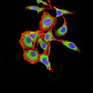
Figure 5:Immunofluorescence analysis of SK-OV-3 cells using ARF1 mouse mAb (green). Blue: DRAQ5 fluorescent DNA dye. Red: Actin filaments have been labeled with Alexa Fluor- 555 phalloidin. Secondary antibody from Fisher (Cat#: 35503)
Flow cytometric
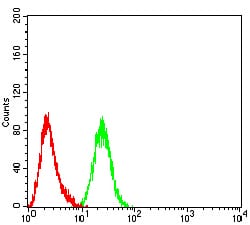
Figure 6:Flow cytometric analysis of Hela cells using ARF1 mouse mAb (green) and negative control (red).
Immunohistochemical analysis
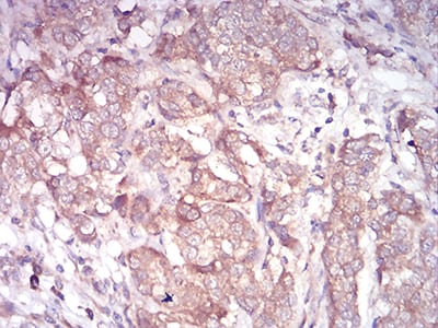
Figure 7:Immunohistochemical analysis of paraffin-embedded bladder cancer tissues using ARF1 mouse mAb with DAB staining.
Immunohistochemical analysis
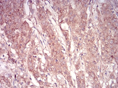
Figure 8:Immunohistochemical analysis of paraffin-embedded stomach cancer tissues using ARF1 mouse mAb with DAB staining.
For Research Use Only. Not for use in diagnostic procedures.