

APP Primary Antibody
Item Information
Catalog #
Size
Price
Description
This gene encodes a cell surface receptor and transmembrane precursor protein that is cleaved by secretases to form a number of peptides. Some of these peptides are secreted and can bind to the acetyltransferase complex APBB1/TIP60 to promote transcriptional activation, while others form the protein basis of the amyloid plaques found in the brains of patients with Alzheimer disease. Mutations in this gene have been implicated in autosomal dominant Alzheimer disease and cerebroarterial amyloidosis (cerebral amyloid angiopathy). Multiple transcript variants encoding several different isoforms have been found for this gene.
Product Overview
Entrez GenelD
351
Aliases
AAA; AD1; PN2; ABPP; APPI; CVAP; ABETA; PN-II; CTFgamma
Clone#
3F12F6
Host / Isotype
Mouse / IgG1
Species Reactivity
Human
Immunogen
Purified recombinant fragment of human APP (AA: 483-699) expressed in E. Coli.
Formulation
Purified antibody in PBS with 0.05% sodium azide.
Storage
Store at 4°C short term. Aliquot and store at -20°C long term. Avoid freeze/thaw cycles.
Product Applications
WB (Western Blot)
1/500 - 1/2000
ICC (Immunocytochemistry)
1/200 - 1/1000
ELISA
1/10000
References
1. J Alzheimers Dis. 2013;35(2):285-95.
2. Proc Natl Acad Sci U S A. 2012 Jul 24;109(30):E2077-82.
2. Proc Natl Acad Sci U S A. 2012 Jul 24;109(30):E2077-82.
Product Image
Western Blot

Figure 1: Western blot analysis using APP mAb against human APP (AA: 483-699) recombinant protein. (Expected MW is 50.7 kDa)
Western Blot

Figure 2: Western blot analysis using APP mAb against HEK293 (1) and APP (AA: 483-699)-hIgGFc transfected HEK293 (2) cell lysate.
Immunofluorescence analysis

Figure 3: Immunofluorescence analysis of A431 cells using APP mouse mAb (green). Blue: DRAQ5 fluorescent DNA dye. Red: Actin filaments have been labeled with Alexa Fluor-555 phalloidin. Secondary antibody from Fisher (Cat#: 35503)
Elisa
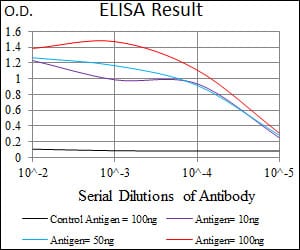
Black line: Control Antigen (100 ng); Purple line: Antigen(10ng); Blue line: Antigen (50 ng); Red line: Antigen (100 ng);
For Research Use Only. Not for use in diagnostic procedures.