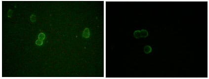
ApoM Primary Antibody

Item Information
Catalog #
Size
Price
Description

ApoM (apolipoprotein M, also designated G3a or NG20), with 188-amino acid protein(about 21kDa), is an apolipoprotein and member of the lipocalin protein family. The Apo-proteins are involved in the specific binding of cellular receptors, the regulation of lipolytic enzymes, and the process of lipid exchange. The encoded protein is secreted through the plasma membrane but remains membrane-bound, where it is involved in lipid transport. The N-terminal region of Apo-M contains hydrophobic residues that may promote association with the phospholipid layer of lipoprotein particles. In vitro, Apo-M is glycosylated when translated in the presence of microsomes, and remains associated with the microsomes after carbonate treatment. Apo-M is expressed in liver and kidney, and is secreted into the bloodstream in HDLs, and also found in triglyceride-rich lipoproteins and LDLs.

Product Overview
Entrez GenelD
55937
Aliases
G3a; NG20; HSPC336; MGC22400
Clone#
10C3G5
Host / Isotype
Mouse / IgG2a
Species Reactivity
Human
Immunogen
Purified recombinant fragment of ApoM expressed in E. Coli.
Formulation
Ascitic fluid containing 0.03% sodium azide.
Storage
4°C; -20°C for long term storage
Product Applications
WB (Western Blot)
1/500 - 1/2000
ICC (Immunocytochemistry)
1/200 - 1/1000
ELISA
1/10000
References
1. Xu,N. & Dahlback,B.1999. J. Biol. Chem. 274 (44):31286-31290.
2. Christian Wolfrum, Matthew N Poy & Markus Stoffel. 2005. Nat Med. 11(4):418-22.
3. Xu,N., Nilsson-Ehle,P. & Ahren,B. 2004. J. Nutr. Biochem. 15 (10):579-582.
4. Zhang,X.Y. ,et al.2004. Acta Histochem. 106 (2):123-128.
Product Image
Western Blot
Figure 1: Western blot analysis using ApoM mouse mAb against GST-ApoM recombinant protein (1) and human serum (2).
Immunofluorescence analysis
Figure 2: Immunofluorescence analysis of methanol-fixed L-02 (left) and Cos7 (right) cells using ApoM mouse mAb showing cytoplasmic and membrane localization.
For Research Use Only. Not for use in diagnostic procedures.