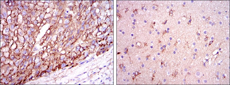
ApoE Primary Antibody

Item Information
Catalog #
Size
Price
Description

Chylomicron remnants and very low density lipoprotein (VLDL) remnants are rapidly removed from the circulation by receptor-mediated endocytosis in the liver. Apolipoprotein E, a main apoprotein of the chylomicron, binds to a specific receptor on liver cells and peripheral cells. ApoE is essential for the normal catabolism of triglyceride-rich lipoprotein constituents. The APOE gene is mapped to chromosome 19 in a cluster with APOC1 and APOC2. Defects in apolipoprotein E result in familial dysbetalipoproteinemia, or type III hyperlipoproteinemia (HLP III), in which increased plasma cholesterol and triglycerides are the consequence of impaired clearance of chylomicron and VLDL remnants. Tissue specificity: Occurs in all lipoprotein fractions in plasma. It constitutes 10-20% of very low density lipoproteins (VLDL) and 1-2% of high density lipoproteins (HDL). APOE is produced in most organs. Significant quantities are produced in liver, brain, spleen, lung, adrenal, ovary, kidney and muscle.

Product Overview
Entrez GenelD
348
Aliases
AD2; LPG; LDLCQ5; MGC1571
Clone#
1H4
Host / Isotype
Mouse / IgG1
Species Reactivity
Human
Immunogen
Purified recombinant fragment of human ApoE expressed in E. Coli.
Formulation
Ascitic fluid containing 0.03% sodium azide.
Storage
4°C; -20°C for long term storage
Product Applications
WB (Western Blot)
1/500 - 1/2000
IHC_P(Immunohistochemistry)
1/200 - 1/1000
FCM (Flow Cytometry)
1/200 - 1/400
ELISA
1/10000
References
1. Arch Dermatol Res. 2009 Jul;301(6):405-10.
2. Pharmacogenomics J. 2009 Aug;9(4):248-57.
Product Image
Western Blot
Figure 1: Western blot analysis using ApoE mAb against HEK293 (1) and ApoE (AA: 20-267)-hIgGFc transfected HEK293 (2) cell lysate.
Immunohistochemical analysis
Figure 2: Immunohistochemical analysis of paraffin-embedded liver cancer tissues (left) and brain tissues (right) using ApoE mouse mAb with DAB staining.
Flow cytometric
Figure 3: Flow cytometric analysis of HepG2 cells using ApoE mouse mAb (green) and negative control (purple).
Elisa
Red: Control Antigen (100ng); Purple: Antigen (10ng); Green: Antigen (50ng); Blue: Antigen (100ng);
For Research Use Only. Not for use in diagnostic procedures.