

APBA2 Primary Antibody
Item Information
Catalog #
Size
Price
Description
The protein encoded by this gene is a member of the X11 protein family. It is a neuronal adapter protein that interacts with the Alzheimer's disease amyloid precursor protein (APP). It stabilizes APP and inhibits production of proteolytic APP fragments including the A beta peptide that is deposited in the brains of Alzheimer's disease patients. This gene product is believed to be involved in signal transduction processes. It is also regarded as a putative vesicular trafficking protein in the brain that can form a complex with the potential to couple synaptic vesicle exocytosis to neuronal cell adhesion. Multiple transcript variants encoding different isoforms have been found for this gene.
Product Overview
Entrez GenelD
321
Aliases
X11L; MINT2; LIN-10; HsT16821; X11-BETA; D15S1518E; MGC:14091
Clone#
7G5G11
Host / Isotype
Mouse / IgG1
Species Reactivity
Human
Immunogen
Purified recombinant fragment of human APBA2 (AA: 15-158) expressed in E. Coli.
Formulation
Purified antibody in PBS with 0.05% sodium azide
Storage
Store at 4°C short term. Aliquot and store at -20°C long term. Avoid freeze/thaw cycles.
Product Applications
WB (Western Blot)
1/500 - 1/2000
FCM (Flow Cytometry)
1/200 - 1/400
ELISA
1/10000
References
1.Neuroreport. 2012 Feb 15;23(3):146-51.
2.Autism Res. 2009 Dec;2(6):359-64.
2.Autism Res. 2009 Dec;2(6):359-64.
Product Image
Elisa

Figure 1: Black line: Control Antigen (100 ng);Purple line: Antigen (10ng); Blue line: Antigen (50 ng); Red line:Antigen (100 ng)
Western Blot

Figure 2:Western blot analysis using APBA2 mAb against human APBA2 (AA: 15-158) recombinant protein. (Expected MW is 42 kDa)
Western Blot
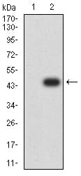
Figure 3:Western blot analysis using APBA2 mAb against HEK293 (1) and APBA2 (AA: 15-158)-hIgGFc transfected HEK293 (2) cell lysate.
Flow cytometric

Figure 4:Flow cytometric analysis of Hela cells using APBA2 mouse mAb (green) and negative control (red).
For Research Use Only. Not for use in diagnostic procedures.