

ANXA1 Primary Antibody
Item Information
Catalog #
Size
Price
Description
Annexin I belongs to a family of Ca(2+)-dependent phospholipid binding proteins which have a molecular weight of approximately 35,000 to 40,000 and are preferentially located on the cytosolic face of the plasma membrane. Annexin I protein has an apparent relative molecular mass of 40 kDa, with phospholipase A2 inhibitory activity. Since phospholipase A2 is required for the biosynthesis of the potent mediators of inflammation, prostaglandins and leukotrienes, annexin I may have potential anti-inflammatory activity.
Product Overview
Entrez GenelD
301
Aliases
ANX1; LPC1
Clone#
2F1
Host / Isotype
Mouse / IgG1
Species Reactivity
Human, Mouse, Monkey
Immunogen
Purified recombinant fragment of human ANXA1 (AA: 144-248 ) expressed in E. Coli.
Formulation
Purified antibody in PBS with 0.05% sodium azide
Storage
Store at 4°C short term. Aliquot and store at -20°C long term. Avoid freeze/thaw cycles.
Product Applications
WB (Western Blot)
1/500 - 1/2000
IHC_P(Immunohistochemistry)
1/200 - 1/1000
ELISA
1/10000
References
1.Cancer Lett. 2010 Aug 1;294(1):111-7. 2.Pathology. 2010 Jan;42(1):43-9.
Product Image
Western Blot

Figure 1: Western blot analysis using ANXA1 mAb against human ANXA1 recombinant protein. (Expected MW is 33.7 kDa)
Immunohistochemical analysis

Figure 2: Immunohistochemical analysis of paraffin-embedded ovarian cancer tissues using ANXA1 mouse mAb with DAB staining.
Western Blot

Figure 2: Western blot analysis using ANXA1 mouse mAb against Hela (1), A549 (2), K562 (3), NIH3T3 (4), C6 (5), and COS7 (6) cell lysate.
Immunohistochemical analysis
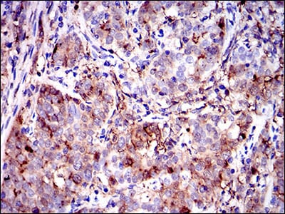
Figure 3: Immunohistochemical analysis of paraffin-embedded cervical cancer tissues using ANXA1 mouse mAb with DAB staining.
Elisa
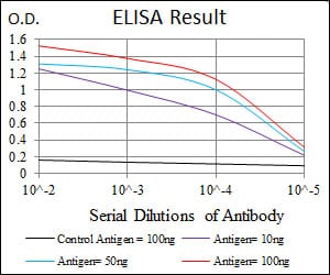
Black line: Control Antigen (100 ng); Purple line: Antigen(10ng); Blue line: Antigen (50 ng); Red line: Antigen (100 ng);
For Research Use Only. Not for use in diagnostic procedures.