

ANAPC10 Primary Antibody
Item Information
Catalog #
Size
Price
Description
ANAPC10 is a core subunit of the anaphase-promoting complex (APC), or cyclosome, a ubiquitin protein ligase that is essential for progression through the cell cycle. APC initiates sister chromatid separation by ubiquitinating the anaphase inhibitor securin (PTTG1; MIM 604147) and triggers exit from mitosis by ubiquitinating cyclin B (CCNB1; MIM 123836), the activating subunit of cyclin-dependent kinase-1 (CDK1; MIM 116940) (summary by Wendt et al., 2001 [PubMed 11524682]).
Product Overview
Entrez GenelD
10393
Aliases
DOC1; APC10
Clone#
8F1D10
Host / Isotype
Mouse / IgG1
Species Reactivity
Human
Immunogen
Purified recombinant fragment of human ANAPC10 (AA: 1-185) expressed in E. Coli.
Formulation
Purified antibody in PBS with 0.05% sodium azide
Storage
Store at 4°C short term. Aliquot and store at -20°C long term. Avoid freeze/thaw cycles.
Product Applications
WB (Western Blot)
1/500 - 1/2000
IHC_P(Immunohistochemistry)
1/200 - 1/1000
ICC (Immunocytochemistry)
1/200 - 1/1000
FCM (Flow Cytometry)
1/200 - 1/400
ELISA
1/10000
References
1.BMC Cell Biol. 2004 May 16;5:20.
2.Nat Struct Biol. 2001 Sep;8(9):784-8.
2.Nat Struct Biol. 2001 Sep;8(9):784-8.
Product Image
Elisa

Figure 1: Black line: Control Antigen (100 ng);Purple line: Antigen (10ng); Blue line: Antigen (50 ng); Red line:Antigen (100 ng)
Western Blot
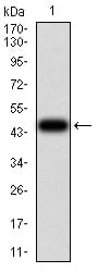
Figure 2:Western blot analysis using ANAPC10 mAb against human ANAPC10 (AA: 1-185) recombinant protein. (Expected MW is 47.2 kDa)
Western Blot

Figure 3:Western blot analysis using ANAPC10 mAb against HEK293 (1) and ANAPC10 (AA: 1-185)-hIgGFc transfected HEK293 (2) cell lysate.
Western Blot

Figure 4:Western blot analysis using ANAPC10 mouse mAb against Hela (1), MCF-7 (2), SK-Br-3 (3), A431 (4), HEK293 (5), A549 (6), and SPC-A-1 (7) cell lysate.
Immunofluorescence analysis

Figure 5:Immunofluorescence analysis of Hela cells using ANAPC10 mouse mAb. Blue: DRAQ5 fluorescent DNA dye. Red: Actin filaments have been labeled with Alexa Fluor- 555 phalloidin.
Immunofluorescence analysis

Figure 6:Immunofluorescence analysis of Hela cells using ANAPC10 mouse mAb (green). Blue: DRAQ5 fluorescent DNA dye. Red: Actin filaments have been labeled with Alexa Fluor- 555 phalloidin. Secondary antibody from Fisher (Cat#: 35503)
Flow cytometric

Figure 7:Flow cytometric analysis of Hela cells using ANAPC10 mouse mAb (green) and negative control (red).
Immunohistochemical analysis

Figure 8:Immunohistochemical analysis of paraffin-embedded colon cancer tissues using ANAPC10 mouse mAb with DAB staining.
Immunohistochemical analysis

Figure 9:Immunohistochemical analysis of paraffin-embedded esophageal cancer tissues using ANAPC10 mouse mAb with DAB staining.
For Research Use Only. Not for use in diagnostic procedures.