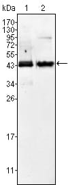
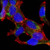
AMACR Primary Antibody

Item Information
Catalog #
Size
Price
Description

AMACR (alpha-methylacyl-CoA racemase) has been recently described as prostate cancer-specific gene that encodes a protein involved in the beta-oxidation of branched chain fatty acids. Expression of AMACR protein is found in prostatic adenocarcinoma but not in benign prostatic tissue. It stains premalignant lesions of prostate: high-grade prostatic intraepithelial neoplasia (PIN) and atypical adenomatous hyperplasia. AMACR can be used as a positive marker for PIN. Defects in AMACR are the cause of congenital bile acid synthesis defect type 4 (CBAS4); also known as cholestasis, intrahepatic, with defective conversion of trihydroxycoprostanic acid to cholic acid or trihydroxycoprostanic acid in bile. Clinical features include neonatal jaundice, intrahepatic cholestasis, bile duct deficiency and absence of cholic acid from bile.

Product Overview
Entrez GenelD
23600
Aliases
RACE
Clone#
2A10F3
Host / Isotype
Mouse / IgG2b
Species Reactivity
Human, Mouse
Immunogen
Purified recombinant fragment of human AMACR expressed in E. Coli.
Formulation
Ascitic fluid containing 0.03% sodium azide.
Storage
4°C; -20°C for long term storage
Product Applications
WB (Western Blot)
1/500 - 1/2000
IHC_P(Immunohistochemistry)
1/200 - 1/1000
ICC (Immunocytochemistry)
1/200 - 1/1000
ELISA
1/10000
References
1. Chen Q. Watson JT. Marengo SR. et al. Cancer Lett. 2006, Dec 8, 244 (2):274-88.Epub 2006 Feb 23.
2. Epstein JI. Herawi M. J Urol. 2006, Mar, 175 (3 Pt 1):820-34. Review.
Product Image
Western Blot
Figure 1: Western blot analysis using AMACR mouse mAb against Jurkat (1) and LNCaP (2) cell lysate.
Immunohistochemical analysis
Figure 2: Immunohistochemical analysis of paraffin-embedded human normal prostate tissues (left) and prostate adenocarcinoma tissues (right), showing cytoplasmic localization using AMACR mouse mAb with DAB staining.
Immunohistochemical analysis
Figure 3: Immunohistochemical analysis of paraffin-embedded human brain cerebellum using AMACR mouse mAb.
Immunofluorescence analysis
Figure 4: Confocal Immunofluorescence analysis of LNCaP cells using AMACR mouse mAb (green). Red: Actin filaments have been labeled with DY-554 phalloidin. Blue: DRAQ5 fluorescent DNA dye.
For Research Use Only. Not for use in diagnostic procedures.