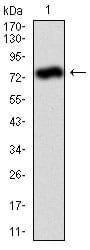
ALPL Primary Antibody

Item Information
Catalog #
Size
Price
Description

There are at least four distinct but related alkaline phosphatases: intestinal, placental, placental-like, and liver/bone/kidney (tissue non-specific). The first three are located together on chromosome 2, while the tissue non-specific form is located on chromosome 1. The product of this gene is a membrane bound glycosylated enzyme that is not expressed in any particular tissue and is, therefore, referred to as the tissue-nonspecific form of the enzyme. The exact physiological function of the alkaline phosphatases is not known. A proposed function of this form of the enzyme is matrix mineralization; however, mice that lack a functional form of this enzyme show normal skeletal development. This enzyme has been linked directly to hypophosphatasia, a disorder that is characterized by hypercalcemia and includes skeletal defects. The character of this disorder can vary, however, depending on the specific mutation since this determines age of onset and severity of symptoms. Alternatively spliced transcript variants have been described.

Product Overview
Formulation
Purified antibody in PBS with 0.05% sodium azide
Storage
4°C; -20°C for long term storage
Product Image
Western Blot
Figure 1: Western blot analysis using ALPL mAb against human ALPL (AA: 18-502) recombinant protein. (Expected MW is 78.9 kDa)
Western Blot
Figure 2: Western blot analysis using ALPL mouse mAb against HeLa (1), and NTERA-2 (4) cell lysate.
Immunohistochemical analysis
Figure 3: Immunohistochemical analysis of paraffin-embedded ovarian cancer tissues using ALPL mouse mAb with DAB staining.
Flow cytometric
Figure 4: Flow cytometric analysis of MCF-7 cells using ALPL mouse mAb (green) and negative control (red).
Elisa
Black line: Control Antigen (100 ng); Purple line: Antigen(10ng); Blue line: Antigen (50 ng); Red line: Antigen (100 ng);
For Research Use Only. Not for use in diagnostic procedures.