
ALK Primary Antibody
Item Information
Catalog #
Size
Price
Description
This gene encodes a receptor tyrosine kinase, which belongs to the insulin receptor superfamily. This protein comprises an extracellular domain, an hydrophobic stretch corresponding to a single pass transmembrane region, and an intracellular kinase domain. It plays an important role in the development of the brain and exerts its effects on specific neurons in the nervous system. This gene has been found to be rearranged, mutated, or amplified in a series of tumours including anaplastic large cell lymphomas, neuroblastoma, and non-small cell lung cancer. The chromosomal rearrangements are the most common genetic alterations in this gene, which result in creation of multiple fusion genes in tumourigenesis, including ALK (chromosome 2)/EML4 (chromosome 2), ALK/RANBP2 (chromosome 2), ALK/ATIC (chromosome 2), ALK/TFG (chromosome 3), ALK/NPM1 (chromosome 5), ALK/SQSTM1 (chromosome 5), ALK/KIF5B (chromosome 10), ALK/CLTC (chromosome 17), ALK/TPM4 (chromosome 19), and ALK/MSN (chromosome X).
Product Overview
Entrez GenelD
238
Aliases
CD246; NBLST3
Clone#
7A11A4
Host / Isotype
Mouse / IgG1
Species Reactivity
Human
Immunogen
Purified recombinant fragment of human ALK (AA: 1366-1468) expressed in E. Coli.
Formulation
Purified antibody in PBS with 0.05% sodium azide
Storage
Store at 4°C short term. Aliquot and store at -20°C long term. Avoid freeze/thaw cycles.
Product Applications
WB (Western Blot)
1/500 - 1/2000
ICC (Immunocytochemistry)
1/50 - 1/250
FCM (Flow Cytometry)
1/200 - 1/400
ELISA
1/10000
References
1.Nature. 2015 Oct 15;526(7573):453-7.
2.Breast Cancer Res. 2015 Sep 17;17:127.
2.Breast Cancer Res. 2015 Sep 17;17:127.
Product Image
Elisa
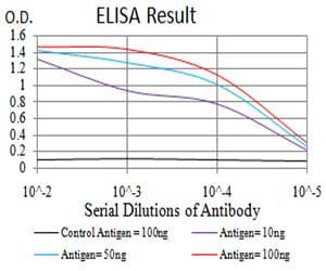
Figure 1: Black line: Control Antigen (100 ng);Purple line: Antigen (10ng); Blue line: Antigen (50 ng); Red line:Antigen (100 ng)
Western Blot
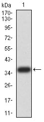
Figure 2:Western blot analysis using ALK mAb against human ALK (AA: 1366-1468) recombinant protein. (Expected MW is 36.9 kDa)
Western Blot
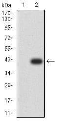
Figure 3:Western blot analysis using ALK mAb against HEK293 (1) and ALK (AA: 1366-1468)-hIgGFc transfected HEK293 (2) cell lysate.
Immunofluorescence analysis
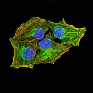
Figure 4:Immunofluorescence analysis of Hela cells using ALK mouse mAb (green). Blue: DRAQ5 fluorescent DNA dye. Red: Actin filaments have been labeled with Alexa Fluor- 555 phalloidin. Secondary antibody from Fisher (Cat#: 35503)
Flow cytometric
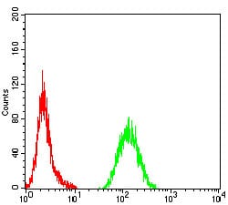
Figure 5:Flow cytometric analysis of Hela cells using ALK mouse mAb (green) and negative control (red).
For Research Use Only. Not for use in diagnostic procedures.