

ALDOA Primary Antibody
Item Information
Catalog #
Size
Price
Description
The protein encoded by this gene, Aldolase A (fructose-bisphosphate aldolase), is a glycolytic enzyme that catalyzes the reversible conversion of fructose-1,6-bisphosphate to glyceraldehyde 3-phosphate and dihydroxyacetone phosphate. Three aldolase isozymes (A, B, and C), encoded by three different genes, are differentially expressed during development. Aldolase A is found in the developing embryo and is produced in even greater amounts in adult muscle. Aldolase A expression is repressed in adult liver, kidney and intestine and similar to aldolase C levels in brain and other nervous tissue. Aldolase A deficiency has been associated with myopathy and hemolytic anemia. Alternative splicing and alternative promoter usage results in multiple transcript variants. Related pseudogenes have been identified on chromosomes 3 and 10.
Product Overview
Entrez GenelD
226
Aliases
ALDA; GSD12; HEL-S-87p
Clone#
2H2B1
Host / Isotype
Mouse / IgG1
Species Reactivity
Human
Immunogen
Purified recombinant fragment of human ALDOA (AA: 9-145) expressed in E. Coli.
Formulation
Purified antibody in PBS with 0.05% sodium azide
Storage
Store at 4°C short term. Aliquot and store at -20°C long term. Avoid freeze/thaw cycles.
Product Applications
WB (Western Blot)
1/500 - 1/2000
ICC (Immunocytochemistry)
1/100 - 1/500
FCM (Flow Cytometry)
1/200 - 1/400
ELISA
1/10000
References
1.Cancer Lett. 2016 Apr 28;374(1):127-35.
2.Oncol Rep. 2014 Nov;32(5):2031-7.
2.Oncol Rep. 2014 Nov;32(5):2031-7.
Product Image
Elisa

Figure 1: Black line: Control Antigen (100 ng);Purple line: Antigen (10ng); Blue line: Antigen (50 ng); Red line:Antigen (100 ng)
Western Blot

Figure 2:Western blot analysis using ALDOA mAb against human ALDOA (AA: 9-145) recombinant protein. (Expected MW is 40.7 kDa)
Western Blot

Figure 3:Western blot analysis using ALDOA mAb against HEK293 (1) and ALDOA (AA: 9-145)-hIgGFc transfected HEK293 (2) cell lysate.
Immunofluorescence analysis
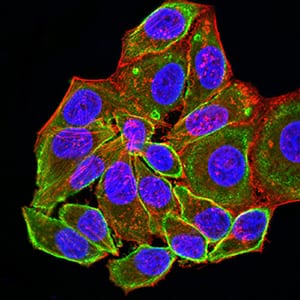
Figure 4:Immunofluorescence analysis of Hela cells using ALDOA mouse mAb (green). Blue: DRAQ5 fluorescent DNA dye. Red: Actin filaments have been labeled with Alexa Fluor- 555 phalloidin. Secondary antibody from Fisher (Cat#: 35503)
Flow cytometric

Figure 5:Flow cytometric analysis of K562 cells using ALDOA mouse mAb (green) and negative control (red).
For Research Use Only. Not for use in diagnostic procedures.