

ALDH1L1 Primary Antibody
Item Information
Catalog #
Size
Price
Description
The protein encoded by this gene catalyzes the conversion of 10-formyltetrahydrofolate, nicotinamide adenine dinucleotide phosphate (NADP+), and water to tetrahydrofolate, NADPH, and carbon dioxide. The encoded protein belongs to the aldehyde dehydrogenase family. Loss of function or expression of this gene is associated with decreased apoptosis, increased cell motility, and cancer progression. There is an antisense transcript that overlaps on the opposite strand with this gene locus. Alternative splicing results in multiple transcript variants.
Product Overview
Entrez GenelD
10840
Aliases
FDH; FTHFD; 10-fTHF; 10-FTHFDH
Clone#
1B5E1
Host / Isotype
Mouse / Mouse IgG1
Species Reactivity
Human, Mouse, Rat
Immunogen
Purified recombinant fragment of human ALDH1L1 (AA: 10-222) expressed in E. Coli.
Formulation
Purified antibody in PBS with 0.05% sodium azide
Storage
Store at 4°C short term. Aliquot and store at -20°C long term. Avoid freeze/thaw cycles.
Product Applications
WB (Western Blot)
1/500 - 1/2000
IHC_P(Immunohistochemistry)
1/200 - 1/1000
FCM (Flow Cytometry)
1/200 - 1/400
ELISA
1/10000
References
1.Chem Biol Interact. 2017 Oct 1;276:23-30.
2.Oncotarget. 2016 Aug 2;7(31):49397-49410.
2.Oncotarget. 2016 Aug 2;7(31):49397-49410.
Product Image
Elisa
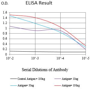
Figure 1:Black line: Control Antigen (100 ng);Purple line: Antigen (10ng); Blue line: Antigen (50 ng); Red line:Antigen (100 ng)
Western Blot

Figure 2:Western blot analysis using ALDH1L1 mAb against human ALDH1L1 (AA: 10-222) recombinant protein. (Expected MW is 26.5 kDa)
Western Blot

Figure 3:Western blot analysis using ALDH1L1 mAb against HEK293-6e (1) and ALDH1L1 (AA: 10-222)-hIgGFc transfected HEK293-6e (2) cell lysate.
Western Blot

Figure 4:Western blot analysis using ALDH1L1 mouse mAb against Rat kidney (1), Mouse liver (2), Rat liver (3) and Mouse kidney (4) tissue lysate.
Immunofluorescence analysis

Figure 5:Flow cytometric analysis of Jurkat cells using ALDH1L1 mouse mAb (green) and negative control (red).
Immunohistochemical analysis

Figure 6:Immunohistochemical analysis of paraffin-embedded liver cancer tissues using ALDH1L1 mouse mAb with DAB staining.
Immunohistochemical analysis

Figure 7:Immunohistochemical analysis of paraffin-embedded bladder cancer tissues using ALDH1L1 mouse mAb with DAB staining.
For Research Use Only. Not for use in diagnostic procedures.