
ALDH1A1 Primary Antibody
Item Information
Catalog #
Size
Price
Description
The protein encoded by this gene belongs to the aldehyde dehydrogenase family. Aldehyde dehydrogenase is the next enzyme after alcohol dehydrogenase in the major pathway of alcohol metabolism. There are two major aldehyde dehydrogenase isozymes in the liver, cytosolic and mitochondrial, which are encoded by distinct genes, and can be distinguished by their electrophoretic mobility, kinetic properties, and subcellular localization. This gene encodes the cytosolic isozyme. Studies in mice show that through its role in retinol metabolism, this gene may also be involved in the regulation of the metabolic responses to high-fat diet.
Product Overview
Entrez GenelD
216
Aliases
ALDC; ALDH1; HEL-9; HEL12; PUMB1; ALDH11; RALDH1; ALDH-E1; HEL-S-53e
Clone#
2B2G1
Host / Isotype
Mouse / IgG1
Species Reactivity
Human
Immunogen
Purified recombinant fragment of human ALDH1A1 (AA: 1-110) expressed in E. Coli.
Formulation
Purified antibody in PBS with 0.05% sodium azide
Storage
Store at 4°C short term. Aliquot and store at -20°C long term. Avoid freeze/thaw cycles.
Product Applications
WB (Western Blot)
1/500 - 1/2000
IHC_P(Immunohistochemistry)
1/200 - 1/1000
FCM (Flow Cytometry)
1/200 - 1/400
ELISA
1/10000
References
1.Oncotarget. 2015 Dec 1;6(38):41360-9.
2.Biomark Med. 2015;9(8):777-90.
2.Biomark Med. 2015;9(8):777-90.
Product Image
Elisa
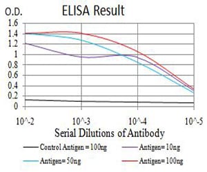
Figure 1: Black line: Control Antigen (100 ng);Purple line: Antigen (10ng); Blue line: Antigen (50 ng); Red line:Antigen (100 ng)
Western Blot
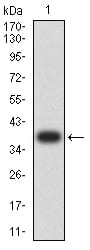
Figure 2:Western blot analysis using ALDH1A1 mAb against human ALDH1A1 (AA: 1-110) recombinant protein. (Expected MW is 38.4 kDa)
Western Blot
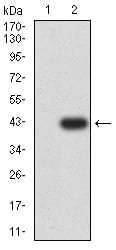
Figure 3:Western blot analysis using ALDH1A1 mAb against HEK293 (1) and ALDH1A1 (AA: 1-110)-hIgGFc transfected HEK293 (2) cell lysate.
Western Blot
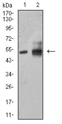
Figure 4:Western blot analysis using ALDH1A1 mouse mAb against HepG2 (1) and A549 (2) cell lysate.
Flow cytometric
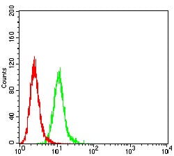
Figure 5:Flow cytometric analysis of HeLa cells using ALDH1A1 mouse mAb (green) and negative control (red).
Immunohistochemical analysis

Figure 6:Immunohistochemical analysis of paraffin-embedded cervical cancer tissues using ALDH1A1 mouse mAb with DAB staining.
Immunohistochemical analysis
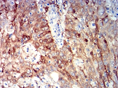
Figure 7:Immunohistochemical analysis of paraffin-embedded stomach cancer tissues using ALDH1A1 mouse mAb with DAB staining.
For Research Use Only. Not for use in diagnostic procedures.