
AKT2 Primary Antibody
Item Information
Catalog #
Size
Price
Description
Akt2 (also designated protein kinase B beta or v-akt murine thymoma viral oncogene homolog 2 ), with 481-amino acid protein (about 53kDa), belongs to the AKT serine/threonine protein kinase family, which also includes Akt1 and Akt3. They are involved in a wide variety of biological processes including cell proliferation, differentiation, apoptosis, tumorigenesis, as well as glycogen synthesis and glucose uptake. Among the members of AKT family, Akt2 is associated with the development of human cancers. Akt2 inhibits cisplatin-induced JNK/p38 and Bax activation through phosphorylation of ASK1 and thus, plays an important role in chemoresistance. Further, Akt2 plays a specific role in muscle differentiation.
Product Overview
Entrez GenelD
208
Aliases
PKBB; PRKBB; PKBBETA; RAC-BETA
Clone#
1B6
Host / Isotype
Mouse / IgG2b
Species Reactivity
Human, Rat, Monkey
Immunogen
Purified recombinant fragment of human AKT2 expressed in E. Coli.
Formulation
Ascitic fluid containing 0.03% sodium azide.
Storage
Store at 4°C short term. Aliquot and store at -20°C long term. Avoid freeze/thaw cycles.
Product Applications
WB (Western Blot)
1/500 - 1/2000
IHC_P(Immunohistochemistry)
1/200 - 1/1000
ICC (Immunocytochemistry)
1/200 - 1/1000
ELISA
1/10000
References
1. Am J Physiol Endocrinol Metab. 2004 Jul;287(1):E8-E15.
2. Oncol Rep. 2004 Jan;11(1):25-32.
3. Cancer Res. 2003 Jan 1;63(1):196-206.
2. Oncol Rep. 2004 Jan;11(1):25-32.
3. Cancer Res. 2003 Jan 1;63(1):196-206.
Product Image
Western Blot
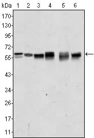
Figure 1: Western blot analysis using AKT2 mouse mAb against A431 (1), Jurkat (2), HEK293 (3), A549 (4), MCF-7 (5) and PC-12 (6) cell lysate.
Immunohistochemical analysis

Figure 2: Immunohistochemical analysis of paraffin-embedded human Thymus tissues using anti-AKT2 mAb
Immunofluorescence analysis
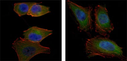
Figure 3: Immunofluorescence analysis of PANC-1 (left) and Hela (right) cells using AKT2 mouse mAb (green). Blue: DRAQ5 fluorescent DNA dye. Red: Actin filaments have been labeled with Alexa Fluor-555 phalloidin.
For Research Use Only. Not for use in diagnostic procedures.