

AGR2 Primary Antibody
Item Information
Catalog #
Size
Price
Description
AGR2 (Anterior Gradient 2, Protein Disulphide Isomerase Family Member) is a Protein Coding gene. Diseases associated with AGR2 include pancreatic ductal adenocarcinoma. Among its related pathways are Tyrosine Kinases / Adaptors and Adhesion. GO annotations related to this gene include dystroglycan binding. An important paralog of this gene is TXNDC12.
Product Overview
Entrez GenelD
10551
Aliases
AG2; GOB-4; HAG-2; XAG-2; PDIA17; HEL-S-116
Clone#
5G1F8
Host / Isotype
Mouse / IgG1
Species Reactivity
Human
Immunogen
Purified recombinant fragment of human AGR2 (AA: 21-175) expressed in E. Coli.
Formulation
Purified antibody in PBS with 0.05% sodium azide
Storage
Store at 4°C short term. Aliquot and store at -20°C long term. Avoid freeze/thaw cycles.
Product Applications
WB (Western Blot)
1/500 - 1/2000
IHC_P(Immunohistochemistry)
1/200 - 1/1000
FCM (Flow Cytometry)
1/200 - 1/400
ELISA
1/10000
References
1.Cancer Sci. 2015 Aug;106(8):1041-9.
2.BMC Cancer. 2014 Nov 3;14:804.
2.BMC Cancer. 2014 Nov 3;14:804.
Product Image
Elisa
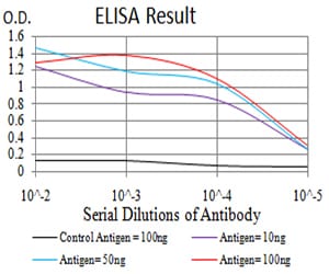
Figure 1:Black line: Control Antigen (100 ng);Purple line: Antigen (10ng); Blue line: Antigen (50 ng); Red line:Antigen (100 ng)
Western Blot
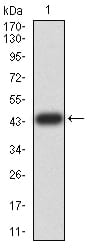
Figure 2:Western blot analysis using AGR2 mAb against human AGR2 (AA: 21-175) recombinant protein. (Expected MW is 43.8 kDa)
Western Blot
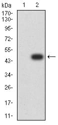
Figure 3:Western blot analysis using AGR2 mAb against HEK293 (1) and AGR2 (AA: 21-175)-hIgGFc transfected HEK293 (2) cell lysate.
Flow cytometric

Figure 4:Flow cytometric analysis of MCF-7 cells using AGR2 mouse mAb (green) and negative control (red).
Immunohistochemical analysis
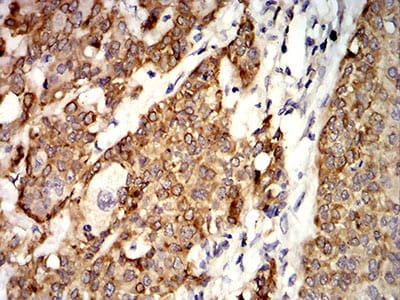
Figure 5:Immunohistochemical analysis of paraffin-embedded bladder cancer tissues using AGR2 mouse mAb with DAB staining.
Immunohistochemical analysis
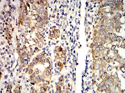
Figure 6:Immunohistochemical analysis of paraffin-embedded stomach cancer tissues using AGR2 mouse mAb with DAB staining.
For Research Use Only. Not for use in diagnostic procedures.