

ADAR Primary Antibody
Item Information
Catalog #
Size
Price
Description
This gene encodes the enzyme responsible for RNA editing by site-specific deamination of adenosines. This enzyme destabilizes double-stranded RNA through conversion of adenosine to inosine. Mutations in this gene have been associated with dyschromatosis symmetrica hereditaria. Alternative splicing results in multiple transcript variants.
Product Overview
Entrez GenelD
103
Aliases
DSH; AGS6; G1P1; IFI4; P136; ADAR1; DRADA; DSRAD; IFI-4; K88DSRBP
Clone#
4E2E4
Host / Isotype
Mouse / IgG1
Species Reactivity
Human
Immunogen
Purified recombinant fragment of human ADAR (AA: 1085-1223) expressed in E. Coli.
Formulation
Purified antibody in PBS with 0.05% sodium azide
Storage
Store at 4°C short term. Aliquot and store at -20°C long term. Avoid freeze/thaw cycles.
Product Applications
WB (Western Blot)
1/500 - 1/2000
ICC (Immunocytochemistry)
1/200 - 1/1000
FCM (Flow Cytometry)
1/200 - 1/400
ELISA
1/10000
References
1.Cell Res. 2015 Apr;25(4):459-76.
2.PLoS One. 2014 Oct 1;9(10):e108476.
2.PLoS One. 2014 Oct 1;9(10):e108476.
Product Image
Elisa
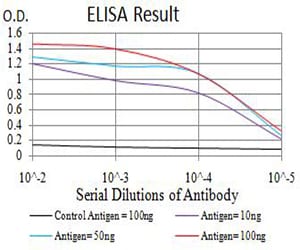
Figure 1: Black line: Control Antigen (100 ng);Purple line: Antigen (10ng); Blue line: Antigen (50 ng); Red line:Antigen (100 ng)
Western Blot
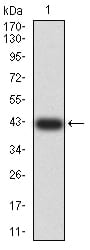
Figure 2:Western blot analysis using ADAR mAb against human ADAR (AA: 1085-1223) recombinant protein. (Expected MW is 42.1 kDa)
Western Blot
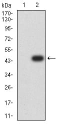
Figure 3:Western blot analysis using ADAR mAb against HEK293 (1) and ADAR (AA: 1085-1223)-hIgGFc transfected HEK293 (2) cell lysate.
Immunofluorescence analysis
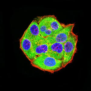
Figure 4:Immunofluorescence analysis of Hela cells using ADAR mouse mAb (green). Blue: DRAQ5 fluorescent DNA dye. Red: Actin filaments have been labeled with Alexa Fluor- 555 phalloidin. Secondary antibody from Fisher (Cat#: 35503)
Flow cytometric
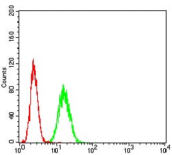
Figure 5:Flow cytometric analysis of Hela cells using ADAR mouse mAb (green) and negative control (red).
Flow cytometric
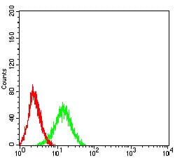
Figure 6:Flow cytometric analysis of Jurkat cells using ADAR mouse mAb (green) and negative control (red).
For Research Use Only. Not for use in diagnostic procedures.