

ACVR1 Primary Antibody
Item Information
Catalog #
Size
Price
Description
Activins are dimeric growth and differentiation factors which belong to the transforming growth factor-beta (TGF-beta) superfamily of structurally related signaling proteins. Activins signal through a heteromeric complex of receptor serine kinases which include at least two type I ( I and IB) and two type II (II and IIB) receptors. These receptors are all transmembrane proteins, composed of a ligand-binding extracellular domain with cysteine-rich region, a transmembrane domain, and a cytoplasmic domain with predicted serine/threonine specificity. Type I receptors are essential for signaling; and type II receptors are required for binding ligands and for expression of type I receptors. Type I and II receptors form a stable complex after ligand binding, resulting in phosphorylation of type I receptors by type II receptors. This gene encodes activin A type I receptor which signals a particular transcriptional response in concert with activin type II receptors. Mutations in this gene are associated with fibrodysplasia ossificans progressive.
Product Overview
Entrez GenelD
90
Aliases
FOP; ALK2; SKR1; TSRI; ACTRI; ACVR1A; ACVRLK2
Clone#
2E2C11
Host / Isotype
Mouse / IgG1
Species Reactivity
Human
Immunogen
Purified recombinant fragment of human ACVR1 (AA: 21-120) expressed in E. Coli.
Formulation
Purified antibody in PBS with 0.05% sodium azide
Storage
Store at 4°C short term. Aliquot and store at -20°C long term. Avoid freeze/thaw cycles.
Product Applications
WB (Western Blot)
1/500 - 1/2000
IHC_P(Immunohistochemistry)
1/200 - 1/1000
ICC (Immunocytochemistry)
1/200 - 1/1000
FCM (Flow Cytometry)
1/200 - 1/400
ELISA
1/10000
References
1.Indian J Pediatr. 2014 Jun;81(6):617-9.
2.Nat Genet. 2014 May;46(5):457-61.
2.Nat Genet. 2014 May;46(5):457-61.
Product Image
Elisa
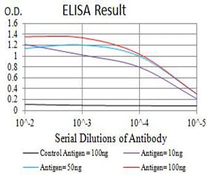
Figure 1: Black line: Control Antigen (100 ng);Purple line: Antigen (10ng); Blue line: Antigen (50 ng); Red line:Antigen (100 ng)
Western Blot
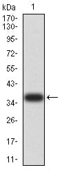
Figure 2:Western blot analysis using ACVR1 mAb against human ACVR1 (AA: 21-120) recombinant protein. (Expected MW is 37.1 kDa)
Western Blot
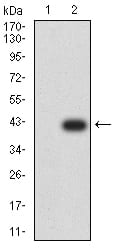
Figure 3:Western blot analysis using ACVR1 mAb against HEK293 (1) and ACVR1 (AA: 21-120)-hIgGFc transfected HEK293 (2) cell lysate.
Immunofluorescence analysis

Figure 4:Immunofluorescence analysis of Hela cells using ACVR1 mouse mAb. Blue: DRAQ5 fluorescent DNA dye. Red: Actin filaments have been labeled with Alexa Fluor- 555 phalloidin.
Immunofluorescence analysis
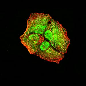
Figure 5:Immunofluorescence analysis of Hela cells using ACVR1 mouse mAb (green). Blue: DRAQ5 fluorescent DNA dye. Red: Actin filaments have been labeled with Alexa Fluor- 555 phalloidin. Secondary antibody from Fisher (Cat#: 35503)
Flow cytometric
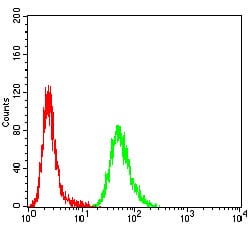
Figure 6:Flow cytometric analysis of Hela cells using ACVR1 mouse mAb (green) and negative control (red).
Immunohistochemical analysis
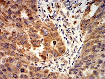
Figure 7:Immunohistochemical analysis of paraffin-embedded ovarian cancer tissues using ACVR1 mouse mAb with DAB staining.
Immunohistochemical analysis
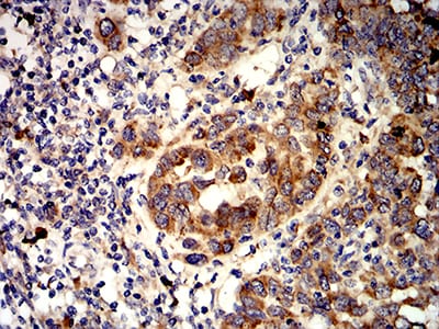
Figure 8:Immunohistochemical analysis of paraffin-embedded endometrial cancer tissues using ACVR1 mouse mAb with DAB staining.
For Research Use Only. Not for use in diagnostic procedures.