ACHE

Item Information
Catalog #
Size
Price
Description

Acetylcholinesterase hydrolyzes the neurotransmitter, acetylcholine at neuromuscular junctions and brain cholinergic synapses, and thus terminates signal transmission. It is also found on the red blood cell membranes, where it constitutes the Yt blood group antigen. Acetylcholinesterase exists in multiple molecular forms which possess similar catalytic properties, but differ in their oligomeric assembly and mode of cell attachment to the cell surface. It is encoded by the single ACHE gene, and the structural diversity in the gene products arises from alternative mRNA splicing, and post-translational associations of catalytic and structural subunits. The major form of acetylcholinesterase found in brain, muscle and other tissues is the hydrophilic species, which forms disulfide-linked oligomers with collagenous, or lipid-containing structural subunits. The other, alternatively spliced form, expressed primarily in the erythroid tissues, differs at the C-terminal end, and contains a cleavable hydrophobic peptide with a GPI-anchor site. It associates with the membranes through the phosphoinositide (PI) moieties added post-translationally. AChE activity may constitute a sensitive biomarker of RBC ageing in vivo, and thus, may be of aid in understanding the effects of transfusion.

Product Overview
Entrez GenelD
43
Aliases
YT; ACEE; ARACHE; N-ACHE
Clone#
3D5E8
Host / Isotype
Mouse / Mouse IgG1
Species Reactivity
Human, Mouse, Monkey, Rat
Immunogen
Purified recombinant fragment of human ACHE expressed in E. Coli.
Formulation
Purified antibody in PBS with 0.05% sodium azide
Storage
4℃; -20℃ for long term storage
Product Applications
WB (Western Blot)
1/500 - 1/2000
IHC_P(Immunohistochemistry)
1/200 - 1/1000
FCM (Flow Cytometry)
1/200 - 1/400
References
1.Molecules. 2020 Oct 4;25(19):4545.
2.J Cell Biochem. 2021 Aug;122(8):787-800.
Product Image
Western Blot
Figure 1:Western blot analysis using ACHE mouse mAb against PC-12 (1), Hela (2),mouse brai (3),NIH/3T3 (4),COS7 (5),Jurkat (6) and Raji (7) cell lysate.
Immunofluorescence analysis
Figure 2:Flow cytometric analysis of NIH/3T3 cells using ACHE mouse mAb (green) and negative control (red).
Immunohistochemical analysis
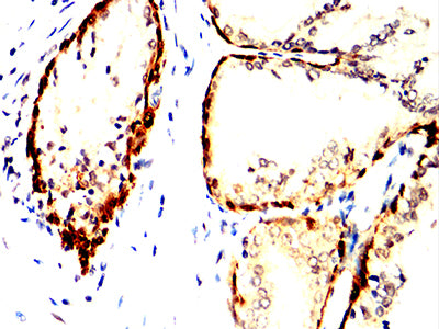
Figure 3:Immunohistochemical analysis of paraffin-embedded prostate cancer tissues using ACHE mouse mAb with DAB staining.
Immunohistochemical analysis
Figure 4:Immunohistochemical analysis of paraffin-embedded rectum cancer tissues using ACHE mouse mAb with DAB staining.
For Research Use Only. Not for use in diagnostic procedures.