

ACAA1 Primary Antibody
Item Information
Catalog #
Size
Price
Description
This gene encodes an enzyme operative in the beta-oxidation system of the peroxisomes. Deficiency of this enzyme leads to pseudo-Zellweger syndrome. Alternative splicing results in multiple transcript variants.
Product Overview
Entrez GenelD
30
Aliases
ACAA; THIO; PTHIO
Clone#
8E7D11
Host / Isotype
Mouse / Mouse IgG1
Species Reactivity
Human, Mouse
Immunogen
Purified recombinant fragment of human ACAA1(AA: 217-315) expressed in E. Coli.
Formulation
Purified antibody in PBS with 0.05% sodium azide
Storage
Store at 4°C short term. Aliquot and store at -20°C long term. Avoid freeze/thaw cycles.
Product Applications
WB (Western Blot)
1/500 - 1/2000
IHC_P(Immunohistochemistry)
1/200 - 1/1000
FCM (Flow Cytometry)
1/200 - 1/400
ELISA
1/10000
References
1.BMC Med Genet. 2011 Dec 8;12:158.
2.J Biol Chem. 2001 Aug 24;276(34):31521-7.
2.J Biol Chem. 2001 Aug 24;276(34):31521-7.
Product Image
Elisa

Figure 1:Black line: Control Antigen (100 ng);Purple line: Antigen (10ng); Blue line: Antigen (50 ng); Red line:Antigen (100 ng)
Western Blot
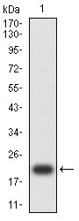
Figure 2:Western blot analysis using ACAA1 mAb against human ACAA1 (AA: 217-315) recombinant protein. (Expected MW is 23.8 kDa)
Western Blot

Figure 3:Western blot analysis using ACAA1 mAb against HEK293-6e (1) and ACAA1 (AA: 217-315)-hIgGFc transfected HEK293-6e (2) cell lysate.
Western Blot
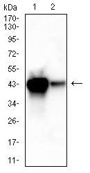
Figure 4:Western blot analysis using ACAA1 mouse mAb against mouse liver (1)and mouse kidney (2) lysate.
Immunofluorescence analysis

Figure 5:Flow cytometric analysis of HL-60 cells using ACAA1 mouse mAb (green) and negative control (red).
Immunohistochemical analysis

Figure 6:Immunohistochemical analysis of paraffin-embedded ovarian cancer tissues using ACAA1 mouse mAb with DAB staining.
Immunohistochemical analysis
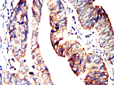
Figure 7:Immunohistochemical analysis of paraffin-embedded rectum cancer tissues using ACAA1 mouse mAb with DAB staining.
For Research Use Only. Not for use in diagnostic procedures.