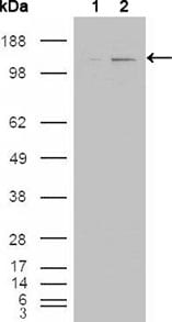
ABL2 Primary Antibody

Item Information
Catalog #
Size
Price
Description

ABL2 (ARG, Abl-related gene), together with c-Abl, forms the Abl family of mammalian non-receptor tyrosine kinases. ABL2 and c-Abl share 89%, 90 and 93% identity in their SH3, SH2 and tyrosine domain, but only 29% identity in the carboxy-terminal half. The human c-Abl and ABL2 genes are expressed ubiquitously. ABL2 had been detected predominantly in the cytoplasm, whereas c-Abl shows both cytoplasmic and nuclear localization. c-Abl is involved in two different chromosomal translocations present in human leukemias, which generate Bcr-Abl and TEL-Abl. Recently, TEL-ARG fusion transcripts have also been identified in acute myeloid leukemias (AML). The Abl family kinases may also interact with receptor tyrosine signaling pathways and regulate cellular function such as cell cycle progression, gene transcription and organization of the actin cytoskeletons in neurons.

Product Overview
Entrez GenelD
27
Aliases
ARG; ABLL; FLJ22224; FLJ31718; FLJ41441
Clone#
1H1
Host / Isotype
Mouse / IgG1
Species Reactivity
Human, Mouse
Immunogen
Purified recombinant fragment of ABL2 expressed in E. Coli.
Formulation
Ascitic fluid containing 0.03% sodium azide.
Storage
4°C; -20°C for long term storage
Product Applications
WB (Western Blot)
1/500 - 1/2000
ICC (Immunocytochemistry)
1/200 - 1/1000
ELISA
1/10000
References
1. Yoshimi I, Takashi I, Tsuneyuki O, et al. Blood. 2000; 95(6): 2126-2131.
2. Scheijen, B. and Griffin, J.D. Oncogene. 2002); 21:3314-33.
Product Image
Western Blot
Figure 1: Western blot analysis using ABL2 mouse mAb against HEK293T cells transfected with the pCMV6-ENTRY control (1) and pCMV6-ENTRY ABL2 cDNA (2).
Immunofluorescence analysis
Figure 2: Immunofluorescence analysis of NIH/3T3 cells using ABL2 mouse mAb (green). Blue: DRAQ5 fluorescent DNA dye. Red: Actin filaments have been labeled with Alexa Fluor-555 phalloidin.
For Research Use Only. Not for use in diagnostic procedures.