

ABCC4 Primary Antibody
Item Information
Catalog #
Size
Price
Description
The protein encoded by this gene is a member of the superfamily of ATP-binding cassette (ABC) transporters. ABC proteins transport various molecules across extra- and intra-cellular membranes. ABC genes are divided into seven distinct subfamilies (ABC1, MDR/TAP, MRP, ALD, OABP, GCN20, White). This protein is a member of the MRP subfamily which is involved in multi-drug resistance. The specific function of this protein has not yet been determined; however, this protein may play a role in cellular detoxification as a pump for its substrate, organic anions. Alternative splicing results in multiple splice variants encoding different isoforms.
Product Overview
Entrez GenelD
10257
Aliases
MRP4; MOATB; MOAT-B; EST170205
Clone#
6A7H3
Host / Isotype
Mouse / IgG1
Species Reactivity
Human
Immunogen
Purified recombinant fragment of human ABCC4 (AA: 631-692) expressed in E. Coli.
Formulation
Purified antibody in PBS with 0.05% sodium azide
Storage
Store at 4°C short term. Aliquot and store at -20°C long term. Avoid freeze/thaw cycles.
Product Applications
WB (Western Blot)
1/500 - 1/2000
IHC_P(Immunohistochemistry)
1/200 - 1/1000
FCM (Flow Cytometry)
1/200 - 1/400
ELISA
1/10000
References
1. Biochem Pharmacol. 2012 Aug 1;84(3):366-73.
2. Arch Dermatol Res. 2012 Jan;304(1):57-63.
2. Arch Dermatol Res. 2012 Jan;304(1):57-63.
Product Image
Western Blot

Figure 1: Western blot analysis using ABCC4 mAb against human ABCC4 recombinant protein. (Expected MW is 32.4 kDa)
Western Blot

Figure 2: Western blot analysis using ABCC4 mAb against HEK293 (1) and ABCC4 (AA: 631-692)-hIgGFc transfected HEK293 (2) cell lysate.
Flow cytometric

Figure 3: Flow cytometric analysis of A549 cells using ABCC4 mouse mAb (green) and negative control (red).
Immunohistochemical analysis
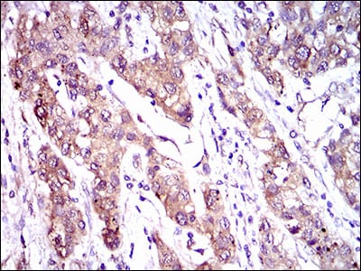
Figure 4: Immunohistochemical analysis of paraffin-embedded liver cancer tissues using ABCC4 mouse mAb with DAB staining.
Immunohistochemical analysis

Figure 5: Immunohistochemical analysis of paraffin-embedded esophagus cancer tissues using ABCC4 mouse mAb with DAB staining.
Elisa

Black line: Control Antigen (100 ng); Purple line: Antigen(10ng); Blue line: Antigen (50 ng); Red line: Antigen (100 ng);
For Research Use Only. Not for use in diagnostic procedures.