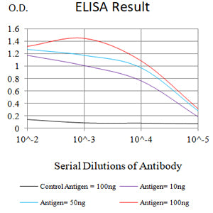
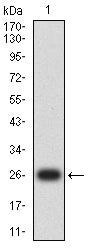
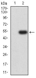
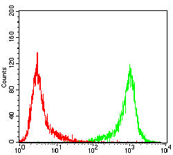
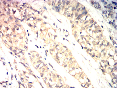
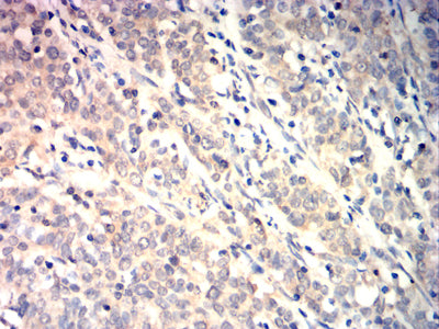
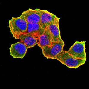
Mouse Monoclonal Antibody to TRIB2

Item Information
Catalog #
Size
Price
Description

This gene encodes one of three members of the Tribbles family. The Tribbles members share a Trb domain, which is homologous to protein serine-threonine kinases, but lacks the active site lysine and probably lacks a catalytic function. The Tribbles proteins interact and modulate the activity of signal transduction pathways in a number of physiological and pathological processes. This Tribbles member induces apoptosis of cells mainly of the hematopoietic origin. It has been identified as a protein up-regulated by inflammatory stimuli in myeloid (THP-1) cells, and also as an oncogene that inactivates the transcription factor C/EBPalpha (CCAAT/enhancer-binding protein alpha) and causes acute myelogenous leukemia. Alternatively spliced transcript variants have been found for this gene. [provided by RefSeq, Mar 2009]

Product Overview
Entrez GenelD
28951
Aliases
C5FW; TRB2; GS3955
Clone#
6A12A12
Host / Isotype
Mouse / IgG2a
Immunogen
Purified recombinant fragment of human TRIB2 (AA: 1-200) expressed in E. Coli.
Formulation
Purified antibody in PBS with 0.05% sodium azide
Storage
4℃; -20℃ for long term storage
Product Applications
WB (Western Blot)
1/500 - 1/2000
IHC_P(Immunohistochemistry)
1/200 - 1/1000
ICC (Immunocytochemistry)
1/200 - 1/1000
FCM (Flow Cytometry)
1/200 - 1/400
ELISA
1/10000
References
1,Int J Cancer. 2017 Oct 15;141(8):1600-1614. 2,Exp Mol Med. 2017 Nov 24;49(11):e401.
Product Image
Elisa
Figure 1:Black line: Control Antigen (100 ng);Purple line: Antigen (10ng); Blue line: Antigen (50 ng); Red line:Antigen (100 ng)
Western Blot
Figure 2:Western blot analysis using TRIB2 mAb against human TRIB2 (AA: 1-200) recombinant protein. (Expected MW is 25.7 kDa)
Western Blot
Figure 3:Western blot analysis using TRIB2 mAb against HEK293-6e (1) and human TRIB2 (AA: 1-200)-hIgGFc transfected HEK293-6e (2) cell lysate.
Flow cytometric analysis
Figure 4:Flow cytometric analysis of Jurkat cells using TRIB2 mouse mAb (green) and negative control (red).
Immunohistochemical analysis
Figure 5:Immunohistochemical analysis of paraffin-embedded bladder cancer tissues using TRIB2 mouse mAb with DAB staining.
Immunohistochemical analysis
Figure 6:Immunohistochemical analysis of paraffin-embedded cervical cancer tissues using TRIB2 mouse mAb with DAB staining.
Immunofluorescence analysis
Figure 7:Immunofluorescence analysis of Hela cells using TRIB2 mouse mAb (green). Blue: DRAQ5 fluorescent DNA dye. Red: Actin filaments have been labeled with Alexa Fluor- 555 phalloidin. Secondary antibody from Fisher (Cat#: 35503)
For Research Use Only. Not for use in diagnostic procedures.