

TET2 Primary Antibody
Item Information
Catalog #
Size
Price
Description
The protein encoded by this gene is a methylcytosine dioxygenase that catalyzes the conversion of methylcytosine to 5-hydroxymethylcytosine. The encoded protein is involved in myelopoiesis, and defects in this gene have been associated with several myeloproliferative disorders. Two variants encoding different isoforms have been found for this gene.
Product Overview
Entrez GenelD
54790
Aliases
MDS; KIAA1546
Clone#
5H8B3
Host / Isotype
Mouse / Mouse IgG1
Immunogen
Purified recombinant fragment of human TET2 expressed in E. Coli.
Formulation
Purified antibody in PBS with 0.05% sodium azide
Storage
4°C; -20°C for long term storage
Product Applications
WB (Western Blot)
1/500 - 1/2000
FCM (Flow Cytometry)
1/200-1/400
ELISA
1/10000
References
1.Int J Lab Hematol. 2019 Oct;41(5):702-709. 2.Clin Epigenetics. 2019 Mar 27;11(1):54.
Product Image
ELISA
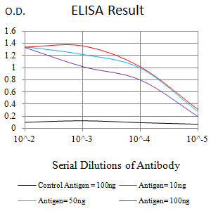
Figure 1: Black line: Control Antigen (100 ng);Purple line: Antigen (10ng); Blue line: Antigen (50 ng); Red line: Antigen (100 ng)
WESTERN BLOT
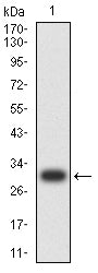
Figure 2: Western blot analysis using TET2 mAb against human TET2 recombinant protein. (Expected MW is 31 kDa)
WESTERN BLOT
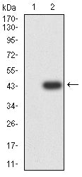
Figure 3: Western blot analysis using TET2 mAb against HEK293 (1) and TET2-hIgGFc transfected HEK293 (2) cell lysate.
FLOW CYTOMETRY
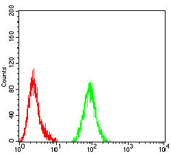
Figure 4: Flow cytometric analysis of Hela cells using TET2 mouse mAb (green) and negative control (red).
For Research Use Only. Not for use in diagnostic procedures.