

SP17 Primary Antibody
Item Information
Catalog #
Size
Price
Description
This gene encodes a protein present at the cell surface. The N-terminus has sequence similarity to human cAMP-dependent protein kinase A (PKA) type II alpha regulatory subunit (RIIa) while the C-terminus has an IQ calmodulin-binding motif. The central portion of the protein has carbohydrate binding motifs and likely functions in cell-cell adhesion. The protein was initially characterized by its involvement in the binding of sperm to the zona pellucida of the oocyte. Recent studies indicate that it is also involved in additional cell-cell adhesion functions such as immune cell migration and metastasis. A retrotransposed pseudogene is present on chromosome 10q22.[provided by RefSeq, Jan 2009]
Product Overview
Entrez GenelD
53340
Aliases
CT22; SPA17; SP17-1
Clone#
1C4C7
Host / Isotype
Mouse / Mouse IgG1
Immunogen
Purified recombinant fragment of human SP17 (AA: 1-152) expressed in E. Coli.
Formulation
Purified antibody in PBS with 0.05% sodium azide
Storage
4°C; -20°C for long term storage
Product Applications
WB (Western Blot)
1/500 - 1/2000
IHC_P(Immunohistochemistry)
1/200-1/1000
FCM (Flow Cytometry)
1/200-1/400
ELISA
1/10000
References
1,Tissue Antigens. 2012 Dec;80(6):523-7.2,BMC Cancer. 2018 Oct 11;18(1):970.3,Cell Immunol. 2015 Nov-Dec;298(1-2):18-24.
Product Image
ELISA
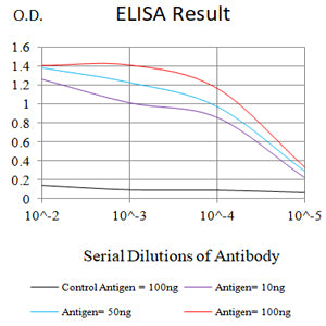
Figure 1: Black line: Control Antigen (100 ng);Purple line: Antigen (10ng); Blue line: Antigen (50 ng); Red line: Antigen (100 ng)
WESTERN BLOT
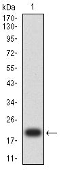
Figure 2: Western blot analysis using SP17 mAb against human SP17 (AA: 1-152) recombinant protein. (Expected MW is 20.3 kDa)
WESTERN BLOT
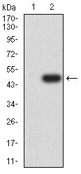
Figure 3: Western blot analysis using SP17 mAb against HEK293-6e (1) and SP17 (AA: 1-152)-hIgGFc transfected HEK293-6e (2) cell lysate.
FLOW CYTOMETRY
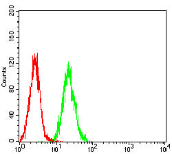
Figure 4: Flow cytometric analysis of SK-OV-3 cells using SP17 mouse mAb (green) and negative control (red).
IMMUNOHISTOCHEMISTRY
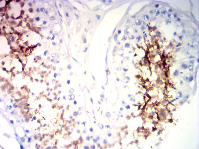
Figure 5: Immunohistochemical analysis of paraffin-embedded testi tissues using SP17 mouse mAb with DAB staining.
IMMUNOHISTOCHEMISTRY
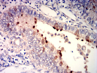
Figure 6: Immunohistochemical analysis of paraffin-embedded endometrial cancer tissues using SP17 mouse mAb with DAB staining.
For Research Use Only. Not for use in diagnostic procedures.