

Mouse Monoclonal Antibody to PSCA
Item Information
Catalog #
Size
Price
Description
This gene encodes a glycosylphosphatidylinositol-anchored cell membrane glycoprotein. In addition to being highly expressed in the prostate it is also expressed in the bladder, placenta, colon, kidney, and stomach. This gene is up-regulated in a large proportion of prostate cancers and is also detected in cancers of the bladder and pancreas. This gene includes a polymorphism that results in an upstream start codon in some individuals; this polymorphism is thought to be associated with a risk for certain gastric and bladder cancers. Alternative splicing results in multiple transcript variants.
Product Overview
Entrez GenelD
8000
Aliases
PRO232
Clone#
3F4E4
Host / Isotype
Mouse / IgG1
Immunogen
Purified recombinant fragment of human PSCA (AA: 1-114) expressed in E. Coli.
Formulation
Purified antibody in PBS with 0.05% sodium azide
Storage
4℃; -20℃ for long term storage
Product Applications
WB (Western Blot)
1/500 - 1/2000
FCM (Flow Cytometry)
1/200 - 1/400
ELISA
1/10000
References
1.Biosci Rep. 2019 Sep 3;39(9):BSR20181025. 2.Cancer Prev Res (Phila). 2019 Sep;12(9):579-584.
Product Image
Elisa
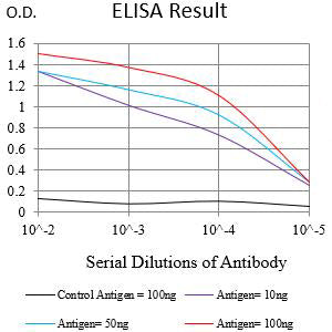
Figure 1:Black line: Control Antigen (100 ng);Purple line: Antigen (10ng); Blue line: Antigen (50 ng); Red line:Antigen (100 ng)
Western Blot
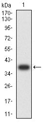
Figure 2:Western blot analysis using PSCA mAb against human PSCA (AA: 1-114) recombinant protein. (Expected MW is 37.8 kDa)
Western Blot
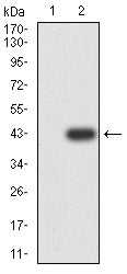
Figure 3:Western blot analysis using PSCA mAb against HEK293-6e (1) and PSCA (AA: 1-114)-hIgGFc transfected HEK293-6e (2) cell lysate.
Flow cytometric analysis
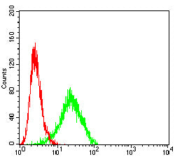
Figure 4:Flow cytometric analysis of PC-3 cells using PSCA mouse mAb (green) and negative control (red).
For Research Use Only. Not for use in diagnostic procedures.