Mouse Monoclonal Antibody to NUP98

Item Information
Catalog #
Size
Price
Description

Nuclear pore complexes (NPCs) regulate the transport of macromolecules between the nucleus and cytoplasm, and are composed of many polypeptide subunits, many of which belong to the nucleoporin family. This gene belongs to the nucleoporin gene family and encodes a 186 kDa precursor protein that undergoes autoproteolytic cleavage to generate a 98 kDa nucleoporin and 96 kDa nucleoporin. The 98 kDa nucleoporin contains a Gly-Leu-Phe-Gly (GLGF) repeat domain and participates in many cellular processes, including nuclear import, nuclear export, mitotic progression, and regulation of gene expression. The 96 kDa nucleoporin is a scaffold component of the NPC. Proteolytic cleavage is important for targeting of the proteins to the NPC. Translocations between this gene and many other partner genes have been observed in different leukemias. Rearrangements typically result in chimeras with the N-terminal GLGF domain of this gene to the C-terminus of the partner gene. Alternative splicing results in multiple transcript variants encoding different isoforms, at least two of which are proteolytically processed. Some variants lack the region that encodes the 96 kDa nucleoporin.
Product Overview
Entrez GenelD
4928
Aliases
ADIR2; NUP96; NUP196; Nup98-96
Clone#
2F5H8
Host / Isotype
Mouse / IgG1
Immunogen
Purified recombinant fragment of human NUP98 (AA: 1-218) expressed in E. Coli.
Formulation
Purified antibody in PBS with 0.05% sodium azide
Storage
4℃; -20℃ for long term storage
Product Applications
WB (Western Blot)
1/500 - 1/2000
FCM (Flow Cytometry)
1/200 - 1/400
ELISA
1/10000
References
1.BMC Cancer. 2019 Apr 2;19(1):236. 2.Nucleus. 2018 Jan 1;9(1):1-8.
Product Image
Elisa
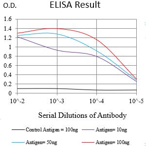
Figure 1:Black line: Control Antigen (100 ng);Purple line: Antigen (10ng); Blue line: Antigen (50 ng); Red line:Antigen (100 ng)
Western Blot
Figure 2:Western blot analysis using NUP98 mAb against human *** (AA: 1-218) recombinant protein. (Expected MW is 31.6 kDa)
Western Blot
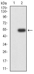
Figure 3:Western blot analysis using NUP98 mAb against HEK293-6e (1) and NUP98 (AA: 1-218)-hIgGFc transfected HEK293-6e (2) cell lysate.
Western Blot
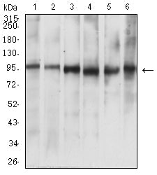
Figure 4:Western blot analysis using NUP98 mouse mAb against A549 (1), L-02 (2), Hela (3), Jurkat (4), HL-60 (5), and COS7 (6) cell lysate.
Flow cytometric analysis
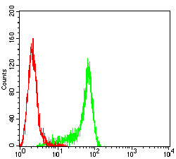
Figure 5:Flow cytometric analysis of Hela cells using NUP98 mouse mAb (green) and negative control (red).
For Research Use Only. Not for use in diagnostic procedures.