

Mouse Monoclonal Antibody to MSH2
Item Information
Catalog #
Size
Price
Description
This locus is frequently mutated in hereditary nonpolyposis colon cancer (HNPCC). When cloned, it was discovered to be a human homolog of the E. coli mismatch repair gene mutS, consistent with the characteristic alterations in microsatellite sequences (RER+ phenotype) found in HNPCC. Two transcript variants encoding different isoforms have been found for this gene.
Product Overview
Entrez GenelD
4436
Aliases
FCC1; COCA1; HNPCC; LCFS2; hMSH2; HNPCC1
Clone#
4D1G12
Host / Isotype
Mouse / IgG1
Immunogen
Purified recombinant fragment of human MSH2 (AA: 2-151) expressed in E. Coli.
Formulation
Purified antibody in PBS with 0.05% sodium azide
Storage
4℃; -20℃ for long term storage
Product Applications
WB (Western Blot)
1/500 - 1/2000
ICC (Immunocytochemistry)
1/200 - 1/1000
FCM (Flow Cytometry)
1/200 - 1/400
ELISA
1/10000
References
1,Genes Chromosomes Cancer. 2020 Feb;59(2):111-118.2,Clin Epigenetics. 2019 Oct 30;11(1):153.
Product Image
Elisa
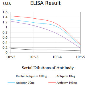
Figure 1:Black line: Control Antigen (100 ng);Purple line: Antigen (10ng); Blue line: Antigen (50 ng); Red line:Antigen (100 ng)
Western Blot
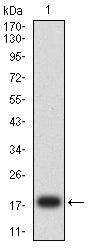
Figure 2:Western blot analysis using MSH2 mAb against human MSH2 (AA: 2-151) recombinant protein. (Expected MW is 19.6 kDa)
Flow cytometric analysis
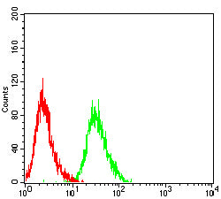
Figure 4:Flow cytometric analysis of Hela cells using MSH2 mouse mAb (green) and negative control (red).
Flow cytometric analysis
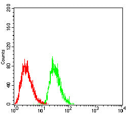
Figure 5:Flow cytometric analysis of HepG2 cells using MSH2 mouse mAb (green) and negative control (red).
Western Blot
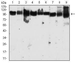
Figure 6:Western blot analysis using MSH2 mouse mAb against MCF-7 (1), A431 (2),K562 (3),Hela (4),Raji (5),A549 (6),NIH/3T3 (7),cos-7 (8), and Hek293-e6 (9) cell lysate.
Immunofluorescence analysis
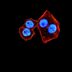
Figure 6:Immunofluorescence analysis of Hela cells using MSH2 mouse mAb (green). Blue: DRAQ5 fluorescent DNA dye. Red: Actin filaments have been labeled with Alexa Fluor- 555 phalloidin. Secondary antibody from Fisher (Cat#: 35503)
For Research Use Only. Not for use in diagnostic procedures.