

Mouse Monoclonal Antibody to HSP70
Item Information
Catalog #
Size
Price
Description
HSPA4 (Heat Shock Protein Family A (Hsp70) Member 4) is a Protein Coding gene. Diseases associated with HSPA4 include Vulvovaginitis and Babesiosis. Among its related pathways are Regulation of degradation of deltaF508 CFTR in CF and Chks in Checkpoint Regulation. An important paralog of this gene is HSPA4L.
Product Overview
Entrez GenelD
3308
Aliases
RY; APG-2; HSPH2; HSPA4; hsp70RY; HEL-S-5a; HS24/P52
Clone#
2E4F10
Host / Isotype
Mouse / IgG1
Immunogen
Purified recombinant fragment of human HSP70 (AA: 642-841) expressed in Mammalian.
Formulation
Purified antibody in PBS with 0.05% sodium azide
Storage
4℃; -20℃ for long term storage
Product Applications
WB (Western Blot)
1/500 - 1/2000
IHC_P(Immunohistochemistry)
1/200 - 1/1000
FCM (Flow Cytometry)
1/200 - 1/400
ELISA
1/10000
References
1,Mol Biol (Mosk). Jan-Feb 2020;54(1):128-136. 2,J Biol Chem. 2020 Jun 12;295(24):8302-8324.
Product Image
Elisa
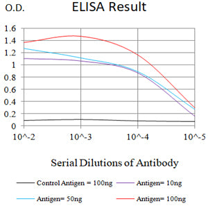
Figure 1:Black line: Control Antigen (100 ng);Purple line: Antigen (10ng); Blue line: Antigen (50 ng); Red line:Antigen (100 ng)
Western Blot
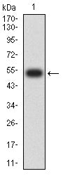
Figure 2:Western blot analysis using HSP70 mAb against human HSP70 (AA: 642-841) recombinant protein. (Expected MW is 53.3 kDa)
Western Blot
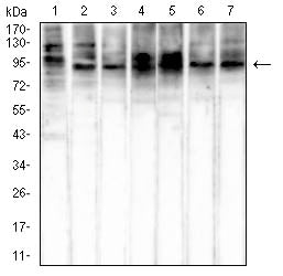
Figure 3:Western blot analysis using HSP70 mouse mAb against NIH/3T3 (1), Hela (2), HepG2 (3), Hek293 (4), COS-7 (5), A549 (6), and Jurkat (7) cell lysate.
Flow cytometric analysis
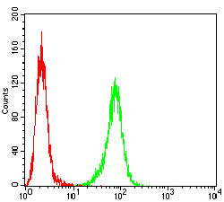
Figure 4:Flow cytometric analysis of Hela cells using HSP70 mouse mAb (green) and negative control (red).
Flow cytometric analysis
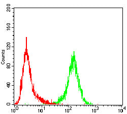
Figure 5:Flow cytometric analysis of Jurkat cells using HSP70 mouse mAb (green) and negative control (red).
Flow cytometric analysis
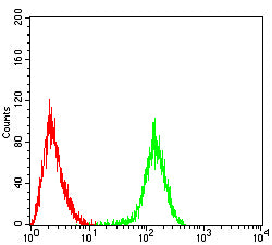
Figure 6:Flow cytometric analysis of Raji cells using HSP70 mouse mAb (green) and negative control (red).
Immunohistochemical analysis
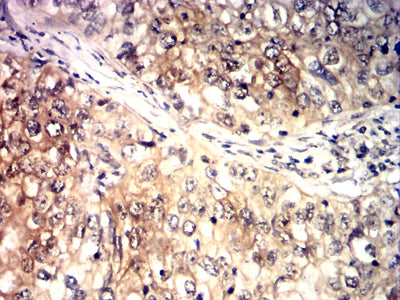
Figure 7:Immunohistochemical analysis of paraffin-embedded breast cancer tissues using HSP70 mouse mAb with DAB staining.
Immunohistochemical analysis
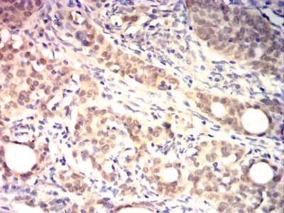
Figure 8:Immunohistochemical analysis of paraffin-embedded cervical cancer tissues using HSP70 mouse mAb with DAB staining.
Immunohistochemical analysis
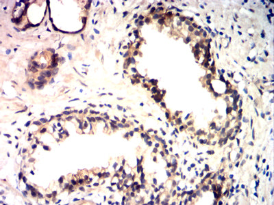
Figure 9:Immunohistochemical analysis of paraffin-embedded prostate cancer tissues using HSP70 mouse mAb with DAB staining.
Immunohistochemical analysis
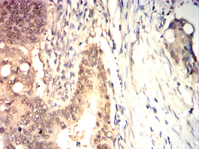
Figure 10:Immunohistochemical analysis of paraffin-embedded rectal cancer tissues using HSP70 mouse mAb with DAB staining.
For Research Use Only. Not for use in diagnostic procedures.