

CHGA Primary Antibody
Item Information
Catalog #
Size
Price
Description
The protein encoded by this gene is a member of the chromogranin/secretogranin family of neuroendocrine secretory proteins. It is found in secretory vesicles of neurons and endocrine cells. This gene product is a precursor to three biologically active peptides; vasostatin, pancreastatin, and parastatin. These peptides act as autocrine or paracrine negative modulators of the neuroendocrine system. Two other peptides, catestatin and chromofungin, have antimicrobial activity and antifungal activity, respectively. Two transcript variants encoding different isoforms have been found for this gene.
Product Overview
Entrez GenelD
1113
Aliases
CGA
Clone#
5H10B12
Host / Isotype
Mouse / Mouse IgG2b
Immunogen
Purified recombinant fragment of human CHGA (AA: 278-457) expressed in E. Coli.
Formulation
Purified antibody in PBS with 0.05% sodium azide
Storage
4°C; -20°C for long term storage
Product Applications
WB (Western Blot)
1/500 - 1/2000
IHC_P(Immunohistochemistry)
1/200-1/1000
FCM (Flow Cytometry)
1/200-1/400
ELISA
1/10000
References
1,Mol Omics. 2019 Feb 11;15(1):67-76;2,Int J Mol Sci. 2019 Jun 14;20(12):2919;3,Pancreas. May/Jun 2019;48(5):662-669.
Product Image
ELISA
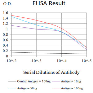
Figure 1: Black line: Control Antigen (100 ng);Purple line: Antigen (10ng); Blue line: Antigen (50 ng); Red line: Antigen (100 ng)
WESTERN BLOT
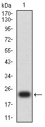
Figure 2: Western blot analysis using CHGA mAb against human CHGA (AA: 278-457) recombinant protein. (Expected MW is 23 kDa)
WESTERN BLOT
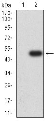
Figure 3: Western blot analysis using CHGA mAb against HEK293-6e (1) and CHGA (AA: 278-457)-hIgGFc transfected HEK293-6e (2) cell lysate.
WESTERN BLOT

Figure 4: Western blot analysis using CHGA mouse mAb against SH-SY5Y (1) cell lysate.
FLOW CYTOMETRY
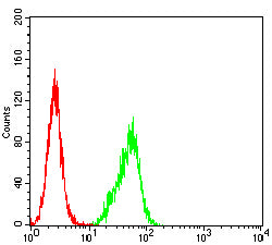
Figure 5: Flow cytometric analysis of C6 cells using CHGA mouse mAb (green) and negative control (red).
FLOW CYTOMETRY

Figure 6: Flow cytometric analysis of Hela cells using CHGA mouse mAb (green) and negative control (red).
IMMUNOHISTOCHEMISTRY
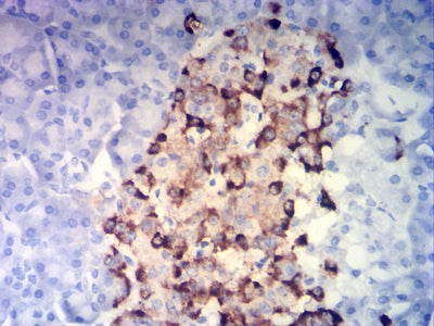
Figure 7: Immunohistochemical analysis of paraffin-embedded pancreatic tissues using CHGA mouse mAb with DAB staining.
For Research Use Only. Not for use in diagnostic procedures.