

Mouse Monoclonal Antibody to CD62E
Item Information
Catalog #
Size
Price
Description
The protein encoded by this gene is found in cytokine-stimulated endothelial cells and is thought to be responsible for the accumulation of blood leukocytes at sites of inflammation by mediating the adhesion of cells to the vascular lining. It exhibits structural features such as the presence of lectin- and EGF-like domains followed by short consensus repeat (SCR) domains that contain 6 conserved cysteine residues. These proteins are part of the selectin family of cell adhesion molecules. Adhesion molecules participate in the interaction between leukocytes and the endothelium and appear to be involved in the pathogenesis of atherosclerosis. [provided by RefSeq, Jul 2008]
Product Overview
Entrez GenelD
6401
Aliases
ELAM; ESEL; SELE; ELAM1; LECAM2
Clone#
5A8C6
Host / Isotype
Mouse / IgG2b
Immunogen
Purified recombinant fragment of human CD62E (AA: extra(22-162)) expressed in E. Coli.
Formulation
Purified antibody in PBS with 0.05% sodium azide
Storage
4℃; -20℃ for long term storage
Product Applications
WB (Western Blot)
1/500 - 1/2000
ICC (Immunocytochemistry)
1/200 - 1/1000
FCM (Flow Cytometry)
1/200 - 1/400
ELISA
1/10000
References
1,PLoS One. 2019 Sep 24;14(9):e0222815. 2,Clin Nephrol. 2020 Jan;93(1):34-49.
Product Image
Elisa
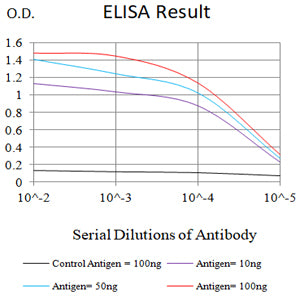
Figure 1:Black line: Control Antigen (100 ng);Purple line: Antigen (10ng); Blue line: Antigen (50 ng); Red line:Antigen (100 ng)
Western Blot
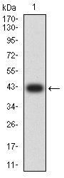
Figure 2:Western blot analysis using CD62E mAb against human CD62E (AA: extra(22-162)) recombinant protein. (Expected MW is 42.3 kDa)
Western Blot
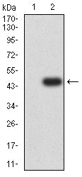
Figure 3:Western blot analysis using CD62E mAb against HEK293-6e (1) and CD62E (AA: extra(22-162))-hIgGFc transfected HEK293-6e (2) cell lysate.
Flow cytometric analysis
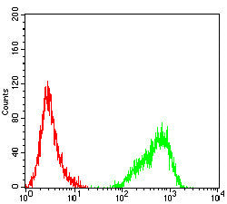
Figure 4:Flow cytometric analysis of Jurkat cells using CD62E mouse mAb (green) and negative control (red).
Immunofluorescence analysis
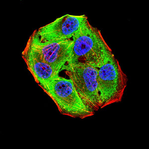
Figure 5:Immunofluorescence analysis of Hela cells using CD62E mouse mAb (green). Blue: DRAQ5 fluorescent DNA dye. Red: Actin filaments have been labeled with Alexa Fluor- 555 phalloidin. Secondary antibody from Fisher (Cat#: 35503)
For Research Use Only. Not for use in diagnostic procedures.