

Mouse Monoclonal Antibody to CD32B
Item Information
Catalog #
Size
Price
Description
The protein encoded by this gene is a low affinity receptor for the Fc region of immunoglobulin gamma complexes. The encoded protein is involved in the phagocytosis of immune complexes and in the regulation of antibody production by B-cells. Variations in this gene may increase susceptibilty to systemic lupus erythematosus (SLE). Several transcript variants encoding different isoforms have been found for this gene. [provided by RefSeq, Jun 2010]
Product Overview
Entrez GenelD
2213
Aliases
FCGR2B;CD32; FCG2; CD32B; FCGR2; IGFR2; FCGR2C; FcRII-c
Clone#
2G6A7
Host / Isotype
Mouse / IgG1
Immunogen
Purified recombinant fragment of human CD32B (AA: 43-217) expressed in E. Coli.
Formulation
Purified antibody in PBS with 0.05% sodium azide
Storage
4℃; -20℃ for long term storage
Product Applications
WB (Western Blot)
1/500 - 1/2000
FCM (Flow Cytometry)
1/200 - 1/400
ELISA
1/10000
References
1,Elife. 2019 Jul 25;8:e46689. 2,J Clin Lab Anal. 2019 Jul;33(6):e22904.
Product Image
Elisa
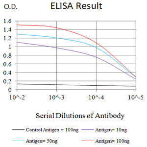
Figure 1:Black line: Control Antigen (100 ng);Purple line: Antigen (10ng); Blue line: Antigen (50 ng); Red line:Antigen (100 ng)
Western Blot
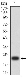
Figure 2:Western blot analysis using CD32B mAb against human CD32B (AA: 43-217) recombinant protein. (Expected MW is 22.5 kDa)
Western Blot
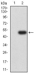
Figure 3:Western blot analysis using CD32B mAb against HEK293-6e (1) and CD32B (AA: 43-217)-hIgGFc transfected HEK293-6e (2) cell lysate.
Flow cytometric analysis
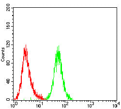
Figure 4:Flow cytometric analysis of Jurkat cells using CD32B mouse mAb (green) and negative control (red).
Flow cytometric analysis
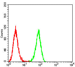
Figure 5:Flow cytometric analysis of THP-1 cells using CD32B mouse mAb (green) and negative control (red).
Flow cytometric analysis
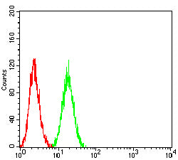
Figure 6:Flow cytometric analysis of K562 cells using CD32B mouse mAb (green) and negative control (red).
For Research Use Only. Not for use in diagnostic procedures.