

Mouse Monoclonal Antibody to CD109
Item Information
Catalog #
Size
Price
Description
This gene encodes a glycosyl phosphatidylinositol (GPI)-linked glycoprotein that localizes to the surface of platelets, activated T-cells, and endothelial cells. The protein binds to and negatively regulates signalling by transforming growth factor beta (TGF-beta). Multiple transcript variants encoding different isoforms have been found for this gene.
Product Overview
Entrez GenelD
135228
Aliases
p180; r150; CPAMD7
Clone#
1A3D3
Host / Isotype
Mouse / IgG1
Immunogen
Purified recombinant fragment of human CD109 (AA: extra 1274-1421) expressed in E. Coli.
Formulation
Purified antibody in PBS with 0.05% sodium azide
Storage
4℃; -20℃ for long term storage
Product Applications
WB (Western Blot)
1/500 - 1/2000
ICC (Immunocytochemistry)
1/20 - 1/100
FCM (Flow Cytometry)
1/200 - 1/400
ELISA
1/10000
References
1.Cancer Sci. 2020 May;111(5):1652-1662. 2.Diagn Pathol. 2015 Aug 7;10:137.
Product Image
Elisa
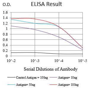
Figure 1:Black line: Control Antigen (100 ng);Purple line: Antigen (10ng); Blue line: Antigen (50 ng); Red line:Antigen (100 ng)
Western Blot
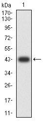
Figure 2:Western blot analysis using CD109 mAb against human CD109 (AA: extra 1274-1421) recombinant protein. (Expected MW is 42.3 kDa)
Western Blot
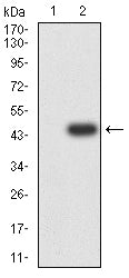
Figure 3:Western blot analysis using CD109 mAb against HEK293-6e (1) and CD109 (AA: extra 1274-1421)-hIgGFc transfected HEK293-6e (2) cell lysate.
Immunofluorescence analysis
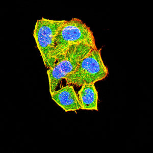
Figure 4:Immunofluorescence analysis of Hela cells using CD109 mouse mAb (green). Blue: DRAQ5 fluorescent DNA dye. Red: Actin filaments have been labeled with Alexa Fluor- 555 phalloidin. Secondary antibody from Fisher (Cat#: 35503)
Flow cytometric analysis
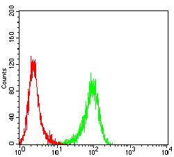
Figure 5:Flow cytometric analysis of Jurkat cells using CD109 mouse mAb (green) and negative control (red).
For Research Use Only. Not for use in diagnostic procedures.